Download
1 / 94
940 likes | 1.26k Vues
Head & Neck Tumours Part II. Dr. Khalid AL-Qahtani MD,MSc,FRCS(c) Assistant Professor Consultant of Otolaryngology Advance Head and Neck Oncology , Thyroid and Parathyroid,Microvascular Reconstruction, Skull Base Surgery. Content. Tumours of the Ears Tumours of the Nose

E N D
Head & Neck TumoursPart II Dr. Khalid AL-Qahtani MD,MSc,FRCS(c) Assistant Professor Consultant of Otolaryngology Advance Head and Neck Oncology , Thyroid and Parathyroid,Microvascular Reconstruction, Skull Base Surgery
Content • Tumours of the Ears • Tumours of the Nose • Tumours of the Mouth • Tumours of the Pharynx • Tumours of the Larynx
Neoplasms of the Ear and Lateral Skull Base • Lesions of the Pinna and EAC • Lesions of the Middle Ear and Mastoid • Lesions of the Petrous Apex and Clivus • Lesions of the IAC, CPA, and Skull Base
Introduction • Generally classified by location, and occasionally by cell-type • Causes of these neoplasms are largely unknown?
Cutaneous carcinoma Squamous cell carcinoma Basal cell carcinoma Malignant melanoma Glandular neoplasm Ceruminous adenoma Ceruminous adenocarcinoma Pleomorphic adenoma Adenoid cystic carcinoma Osteoma and exostosis Miscellaneous neoplasm Merkel cell carcinoma Squamous papilloma Pilomatrixoma Myxoma Auricular endochondrial pseudocyst Chondrodermatitis nodularis chronica helicis (Winkler disease) Neoplasms of the pinna and external auditory canal
Adenomatous neoplasm Benign middle ear adenoma Endolymphatic sac tumor Chordoma Congenital neoplasm Dermoid Teratoma Choristoma Cholesterol granuloma Langerhans cell histiocytosis Eosinophilic granuloma Hand-Schüller-Christian disease Letterer-Siwe disease Sarcoma Rhabdomyosarcoma Chondrosarcoma Ewing sarcoma Osteogenic sarcoma Fibrosarcoma Lesions of the Petrous Apex and Clivus
Neoplasms of the internal auditory canal and cerebellopontine angle Schwannoma Vestibular schwannoma Facial nerve schwannoma Trigeminal schwannoma Jugular foramen schwannomaMeningiomaLipomaMetastases
Neoplasms of the Pinna and EAC Cutaneous Carcinoma BCC • BCC (20% of ear/TB neoplasms) • Most on pinna • Sun exposure is initiator • Locally infiltrative, rolled border central crusting ulcer • May invade TB if left untreated
Cutaneous CarcinomaSCCA • Pinna and EAC are common • Sun, cold, radiation are all factors • Scaly irregular indurated maculopapular lesion, often ulcerated with sero-sang d/c • Can be confused with OE • Other symptoms VII, CHL, SNHL (with invasion of TB) • Met. To LN more common than BCC
Cutaneous CarcinomaTreatment • Moh’s micro surgery for most scc and bcc pinna lesions • TB lesions require TB resection and RT • Address LN in SCC
Osteomata and Exostoses • Benign bony growths in EAC • Osteoma’s – solitary, pedunculated, smooth, round lesions arising from tympanomastoid and squamous suture • Exostose’s – broad, more medial, multiple, often bilateral • Related to cold water exposure
Lesions of the Middle Ear and Mastoid • Paragangliomas • Most common neoplasm of middle ear but still rare • Glomus tympanicum • Originate on promontory of cochlea (jacobson or Arnold’s nerve) • Fill ME space and ossicles involved • May extend to hypotympanum and expose jugular or petrous carotid • Present with HL and pulsatile tinnitus and ME mass • Glomus jugulare • Arise in jugular fossa • Become large before symptomatic (multiple CN)
Lesions of the Middle Ear and Mastoid • Paragangliomas • Brown sign +ve pressure leads to blanching • Aquino sign – ipsilat CA compression decreases pulsation • Vernet syndrome (or JF syndrome) – paresis of CN’s 9, 10, 11 • Villaret Syndrome = JF syndrome plus Horners
Paragangliomas • Rx is complete surgical excision • If secretory must address this (alpha or beta blockade) • Trans canal, trans mastoid-lab, trans cervical, infra temporal, intra cranial • Pre-op embolization is a neccessity • If you think it invades the ICA, balloon occlusion studies must be done • RT or stereotactic radiosurgery can halt disease in up to 90%
Lesions of the Petrous Apex and Clivus • Cholesterol granulomas • Most common lesion of the petrous apex • Negative pressure in lumen causes hemorrhage • Expansile lesion • Hearing loss, tinnitus, vertigo, facial twitching • HRCT • MRI diagnostic • T1 and T2 hyperintense
Cholesterol Granuloma • Causes: poor drainage of ME, hemmorhage, obstruction of ventilation, FB reaction to cholesterol crystals from HB catabolism • Rx is surgical drainage
Lesions of the IAC, CPA, and Skull Base • Schwannomas (no longer acoustic) • Arise from sheaths of cranial nerves • Vestibular, facial, trigeminal, jugular • Varied presentation • HRCT • Inhomogeneous enhancement • Smooth mass effect • MRI – definitive diagnosis • T1- low intensity • Marked enhancement with gadolinium on T1
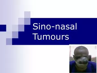
Neoplasms of the Nose and Paranasal Sinus • Introduction • Benign Lesions • Malignant lesions
Neoplasms of Nose and Paranasal Sinuses • Very rare 3% • Delay in diagnosis due to similarity to benign conditions • Nasal cavity • ½ benign • ½ malignant • Paranasal Sinuses • Malignant
Neoplasms of Nose and Paranasal Sinuses • Multimodality treatment • Orbital Preservation • Minimally invasive surgical techniques
Epidemiology • Predominately of older males • Exposure: • Wood, nickel-refining processes • Industrial fumes, leather tanning • Cigarette and Alcohol consumption • No significant association has been shown
Location • Maxillary sinus • 70% • Ethmoid sinus • 20% • Sphenoid • 3% • Frontal • 1%
Presentation • Oral symptoms: 25-35% • Pain, trismus, alveolar ridge fullness, erosion • Nasal findings: 50% • Obstruction, epistaxis, rhinorrhea • Ocular findings: 25% • Epiphora, diplopia, proptosis • Facial signs • Paresthesias, asymmetry
Benign Lesions • Papillomas • Osteomas • Fibrous Dysplasia
Papilloma • Vestibular papillomas • Schneiderian papillomas derived from schneiderian mucosa (squamous) • Fungiform: 50%, nasal septum • Cylindrical: 3%, lateral wall/sinuses • Inverted: 47%, lateral wall
Inverted Papilloma • 4% of sinonasal tumors • Site of Origin: lateral nasal wall • Unilateral • Malignant degeneration in 2-13% (avg 10%)
Inverted PapillomaResection • Initially via transnasal resection: • 50-80% recurrence • Medial Maxillectomy via lateral rhinotomy: • Gold Standard • 10-20% • Endoscopic medial maxillectomy: • Key concepts: • Identify the origin of the papilloma • Bony removal of this region • Recurrent lesions: • Via medial maxillectomy vs. Endoscopic resection • 22%
Osteomas • Benign slow growing tumors of mature bone • Location: • Frontal, ethmoids, maxillary sinuses • When obstructing mucosal flow can lead to mucocele formation • Treatment is local excision
Fibrous dysplasia • Dysplastic transformation of normal bone with collagen, fibroblasts, and osteoid material • Monostotic vs Polyostotic • Surgical excision for obstructing lesions • Malignant transformation to rhabdomyosarcoma has been seen with radiation
Malignant lesions • Squamous cell carcinoma • Adenoid cystic carcinoma • Mucoepidermoid carcinoma • Adenocarcinoma • Hemangiopericytoma • Melanoma • Olfactory neuroblastoma • Osteogenic sarcoma, fibrosarcoma, chondrosarcoma, rhabdomyosarcoma • Lymphoma • Metastatic tumors • Sinonasal undifferentiated carcinoma
Squamous cell carcinoma • Most common tumor (80%) • Location: • Maxillary sinus (70%) • Nasal cavity (20%) • 90% have local invasion by presentation • Lymphatic drainage: • First echelon: retropharyngeal nodes • Second echelon: subdigastric nodes
Staging of Maxillary Sinus Tumors • T1: limited to antral mucosa without bony erosion • T2: erosion or destruction of the infrastructure, including the hard palate and/or middle meatus • T3: Tumor invades: skin of cheek, posterior wall of sinus, inferior or medial wall of orbit, anterior ethmoid sinus • T4: tumor invades orbital contents and/or: cribriform plate, post ethmoids or sphenoid, nasopharynx, soft palate, pterygopalatine or infratemporal fossa or base of skull
Treatment • 88% present in advanced stages (T3/T4) • Surgical resection with postoperative radiation • Complex 3-D anatomy makes margins difficult
Olfactory NeuroblastomaEsthesioneuroblastoma • Originate from stem cells of neural crest origin that differentiate into olfactory sensory cells. • Kadish Classification • A: confined to nasal cavity • B: involving the paranasal cavity • C: extending beyond these limits
Olfactory NeuroblastomaEsthesioneuroblastoma • Aggressive behavior • Local failure: 50-75% • Metastatic disease develops in 20-30% • Treatment: • En bloc surgical resection with postoperative XRT
Oral Cavity Cancer • Introduction • Premalignant Lesions • Malignant Lesions
Epidemiology • 95% are squamous cell carcinoma • Risk factors • Smoking (depends on dosage and type) • Alcohol • Snuff dipping / tobacco chewing • HPV (subtype 16) • Reverse cigar smoking (India) • Betel-nut chewing (Asia) • ?Poor dentition / mechanical irritation (dentures)
Epidemiology • 75% of cases occur on 10% of mucosal surface area • Area from ant FOM along gingivobuccal sulcus and lat border tongue to retromolar trigone and ant tonsil pillar • Flow and pooling of carcinogen-contaminated saliva here • Incidence 4% cancers in males, 2% in females (increasing in females)
Evaluation and Diagnosis • Lesions generally easy to see • Simple biopsy under local anesthesia • Important goals: • Stage full extent of disease • Rule out synchronous primary • Evaluate for possible metastatic disease • CT or MRI for T2 or greater • Staging endoscopy
AJCC TNM Staging • Primary Tumor (T) • Tx: unassessable • T1: tumor 2cm or less in greatest diameter • T2: tumor 2-4cm • T3: tumor >4cm • T4: tumor invades adjacent structures • Cortical bone, deep tongue musculature, maxillary sinus, skin
Differential Diagnosis • Granular cell myoblastoma • Minor salivary gland neoplasm • Adenoid cystic, mucoepidermoid, adeno-ca. • Sarcomas (rhabdo, lipo, MFH, leiomyo) • Hodgkin and NH lymphoma • Malignant melanoma • Hairy leukoplakia, Kaposi sarcoma • HIV, immunocompromised
Premalignant Lesions • Leukoplakia • Hyperkeratosis, dysplasia • Malignant transformation greater in non-smokers • Treatment: • Surgical or laser excision • Topical bleomycin, retinoids, • Erythroplasia • Greater risk of malignancy
Prognostic Factors • Poor prognostic tumor factors include • Tumor thickness (3mm FOM, 5mm tongue) • Stage • Perineural invasion • Lymphatic invasion • Vascular invasion • Neck/distant mets • DNA ploidy • Pathology
Treatment and posttreatment follow-up: neoplasms of the oral cavity SURGERY Primary Resection with adequate margins; frozen section as needed Tracheostomy as needed Feeding tube optional Surgical orientation of specimen for pathologist Neck Modified/radical dissection for unilateral metastatic disease and bilateral dissections for metastases in both necks Suction drainage Perioperative care Antibiotics Hospitalization for 3–10 days Tube feedings Suction drainage for necks(s)—remove when output <25–30 mL/24-h period Suture removal 5–10 days postoperatively
Tumours of Pharynx • Nasopharyngeal Carcinoma • Oropharyngeal Carcinoma • Hypopharyngeal Carcinoma
Nasopharyngeal CarcinomaIntroduction • Rare in the US, more common in Asia • High index of suspicion required for early diagnosis • Nasopharyngeal malignancies • SCCA (“nasopharyngeal carcinoma”) • Lymphoma • Salivary gland tumors • Sarcomas
Classification • WHO classes • Based on light microscopy findings • All SCCA by EM • Type I - “SCCA” • 25 % of NPC (in North Amer population) • 1-2 % NPC of endemic populations • moderate to well differentiated cells similar to other SCCA ( keratin, intercellular bridges)
Classification • Type II - “non-keratinizing” carcinoma • 12 % of NPC • variable differentiation of cells (mature to anaplastic) • minimal if any keratin production • may resemble transitional cell carcinoma of the bladder • Lumped with Type III in 1991 WHO revision