Meninges and CSF
150 likes | 347 Vues
Meninges and CSF. Dr. K. Sivapalan. Meninges. Dura mater, arachnoid mater and pia mater. Dura- single and tough layer of fibrous tissue fused with inner periosteum except in few areas. Arachnoid - fibrocellular tissue. Outer most cells are bonded to each other by tight junctions.

Meninges and CSF
E N D
Presentation Transcript
Meninges and CSF Dr. K. Sivapalan
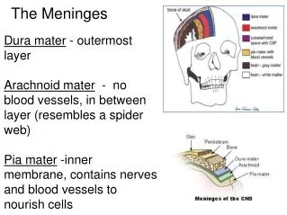
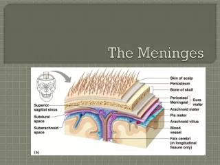
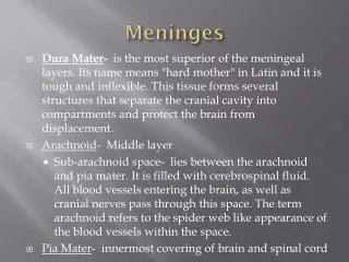
Meninges • Dura mater, arachnoid mater and pia mater. • Dura- single and tough layer of fibrous tissue fused with inner periosteum except in few areas. • Arachnoid- fibrocellular tissue. Outer most cells are bonded to each other by tight junctions. • Subarachnoid space- arachnoidtrabeculae cross the space to reach the pia. [CSF] • Pia- invests the nervous tissue closely lining the outer surface. It and the sub-pial space follow into the tissue with blood vessols. Meningies and CSF
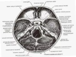
Cranial Dura • The dura is seperated from periosteum only where is is reflected interiorly. • In such places, the intervening space contains venous sinuses. • Falxcerebri- it occupies the longitudinal fissure between the cerebral hemispheres. • Tentoriumcerebelli- it arches like a tent above the posterior cranial fossa between the cerebellum and the cerebrum, lifted by the falxcerebri in the midline Meningies and CSF
Spinal Meninges • Spinal dura is attached to brim of the foramen magneum reaching down to S2 vertibra. • It is attached to the posterior longitudinal ligament of the vertebra in the midline. • Else where it is surrounded by adipose tissue containing internal vertebral plexus of veins. • The dura is pierced by the nerve roots in each segment. The dura follows the nerve root up to the intervertibral foramen. Meningies and CSF
Sheath of the Optic Nerve • The optic nerve is extension of the CNS. • The meningeal covering extends up to the eye around the optic nerve. Meningies and CSF
TentoriumCerebelli • Tentoriumcerebelli is attached to the inner of the occipital bone, along the upper border of the petrous temporal bone, to the posterior clenoid process of the sphenoid bone. • The free margin is U shaped and the mid brain passes through the concavity. • The two limbs projecting forwards are linked by a sheet of dura, diapharagmasellae which covers the pituitary fossae. Meningies and CSF
Innervation of Cranial Dura • Dura of the supra tentorial compartment is innervated by sensory fibers from trigerminal nerve. • Injury or inflamation of supra tentorialmeningies give rise to frontal or parietal head ache. • Dura of the infra tentorial compartment is supplied by upper cervical spinal nerves. • Injury or inflamation of meninges here gives rise to occipital or neck pain and spasm of neck muscles [neck stiffness in meningitis] Meningies and CSF
Subarachnoid Cisterns • Areas of subarachnoid space which contains pools of CSF are the cisterns. • Cisterna magna is the largest between Medulla and the cerebellum. • Cisterna ambiens is on either side of the mid brain. • Cisterna pontis is ventral to the pons. • Inter peduncular cistern is in front of the pons between the cerebral peduncles. Meningies and CSF
Choroid Plexuses • Choroid plexus is found in the 4 ventricles. • Telachoroidea is the double layered fold of pia above the thalami with plexus of vessels forming choroid plexus in two rows in either side. • The middle two are in the roof of the third ventricle and the lateral ones in the floor of the lateral ventricles. • Similar choroid plexus is found in the roof of the fourth ventricle. Meningies and CSF
CSF and ECF in Brain • CSF volume is about 150 ml, production is 550 ml/day, 50-70 % secreted by choroid plexus and the rest by vessels in the walls of the ventricles. • The composition is the same as the ECF in the brain which is 15 % of the brain volume. • The pia is freely permiable to CSF and there is free communication and diffusion of substances between the CSF and the ECF. Meningies and CSF
Composition Meningies and CSF
Circulation of CSF • CSF enters the third ventricle through the inter ventricular foramen. • It descends to the fourth ventricle through the aqueduct. • It enters the subarachnoid space through median and lateral apertures in the roof of the fourth ventricle. • Flow in the central canal of the spinal cord is negligible • Some CSF descends through foramen magnum reaching the lumbar cistern in 12 hours. • Small amount is absorbed into the spinal veins but most returns to subarachnoid space in cranium. • It ascends further through the tentorial notch and around the cerebrum. • Finally it is absorbed into the venous sinuses. Meningies and CSF
Function of the CSF • The weight of brain in air is 1400 g but in the “water bath” of CSF, it is 50 g. • The brain is attached and held in position by arachnoidtrabaculae, blood vessels and nerve roots. Weight reduction by CSF helps the flimsy attachments to hold the brain. • Removal of CSF can cause sever pain because the brain hangs on the vessols. • The brain is protected from the trauma of head injuries by the CSF and meninges. Meningies and CSF
Re-absorption of CSF • CSF is returned to the blood mainly through arachnoid granulations found in the superior sagital sinus and small venous lacunae that open into it. • Arachnoid granulations are pin head pouches of arachnoid matter projecting through the dural wall. • Normal lumbar CSF presure is 70-180 mm CSF [average- 112]. • Secretion and absorption are equal at normal pressure. • Absorption stops below 68 mm CSF. Meningies and CSF
Abnormalities of CSF • When circulation is blocked CSF pressure increases proximal to the obstruction. • Block of foramina in the fourth ventricle or failure of absorption results in hydrocephalus. • Abnormalities in composition signifies the type of disease of the meninges. Meningies and CSF