Real-Time Imaging of Antiproton Annihilation for Medical Applications
This research presents groundbreaking real-time imaging techniques to visualize high-energy gamma rays produced from antiproton annihilation with protons in biological cells. Utilizing the IRIS high-resolution detector at CERN, we demonstrate the potential of antiproton annihilation imaging aimed at cancer treatment at a cellular level. We discuss experimental setups, results from gamma-ray visualization, and the future applications of antiproton technology for precision cancer therapy, highlighting its promise as a novel radiotherapy technique.

Real-Time Imaging of Antiproton Annihilation for Medical Applications
E N D
Presentation Transcript
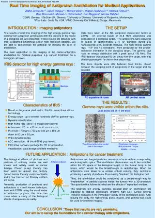
Antiprotons beam Tumor pions gammas muons Presented at EPS 13 (European Physical Society), Bern, Switzerland, July 2005 Real Time Imaging of Antiproton Annihilation for Medical Applications Zlatko Dimcovski1,3, Sylvie Chapuy2,3, Michael Doser1, Dragan Hajdukovic1,4, Mimoza Ristova3,6, Marc Dimcovski2,Michael H. Holzscheiter5, Carl Maggiore5, Nicolas Rabiller2 1CERN, Geneva, 2BioScan SA, Geneva, 3University of Geneva, 4University of Podgorica, Montenegro, 5Pbar Labs, Santa Fe, USA,6PMF, University Kiril &Metodij, Skopje, Macedonia, INTRODUCTION : Imaging antiprotons First results of real time imaging of the high energy gamma rays coming from antiproton annihilation with the protons in the nucleiof a biological cell are presented. These results are obtained with IRIS, a patented amorphous silicon high resolution detector. We are able to demonstrate the potential for imaging the point of annihilation. The main application is the imaging of the proton-antiproton annihilation for medical purposes, e.g. cancer treatment on a biological cell level. THE SETUP Data were taken at the AD, antiproton decelerator facility at CERN. An external beam of 47.8 MeV antiprotons was deposited on a biological target. The antiprotons were delivered in pulses of approximately 3 x 10⁷ particles during 200 nanoseconds at 90 seconds intervals. The high energy gamma rays, ~108 into 4 steradians, were produced by the proton-antiproton interaction in the biological target. The gamma rays have an energy distribution with a peak about 150 MeV. The IRIS detector was placed 50 cm away from the target, with lead shielding protection for the on-line electronics. The tests objects were slits between lead bricks, placed between the stopping point of antiprotons in the target and the detector. IRIS detector for high energy gamma rays : Gamma rays scintillator Antiprotons beam Visible light photons IRIS & beam control systems Test object : slit in lead bricks TFT a-Si photodiode readout device IRIS detector Electrons Real time readout electronics AD experiment room AD control room Digital data output THE RESULTS : Gamma rays were visible within the slits. Characteristics of IRIS: Based on large area pixel matrix, thin film amorphous silicon technology, Energy range : up to several hundreds MeV for gamma-rays, Dynamic visualization, High frame rate : up to 10 images per second, Active area : 20 cm x 20 cm or 41 cm x 41 cm, Pixel size : 750 µm x 750 µm, 400 µm x 400 µm down to 50 µm x 50 µm, Wide dynamic range, ADC resolution : 16 bits (65536 gray levels), IRIS-View, software package for PC for acquisition, visualization, data storage and tele-medecine. Lead bricks slit of ~1 cm thick The slit is visible in the medial part of the image. Distribution histogram. IRIS pitch size750 µm. FWMH : ~11 mm. FUTURE APPLICATION : Antiprotons for cancer treatment The biological effects of photons and particles of ordinary matter are well known and widely used in cancer therapy.Photon (x-ray) therapy has been used for almost one century. Proton cancer therapy exists worldwide and a few heavier ion therapy centers are active. Presently, production of positrons and antiprotons is a well known technique. Now, with CERN being the world leader in antiproton physics, the time has come when a study of the biological effects of antiprotons is reality. Antiprotons, as charged particles, are easy to focus with a corresponding electromagnetic optics. The annihilation phenomenon could be controlled within the 3D space of the biological target, or the human body in the future, which would be the breakthrough in radiotherapy. When the antiprotons slow down to a certain critical velocity, they annihilate, producing a variety of particles, thus making “implose” the biological cell. Thus, the annihilation could be considered as a breakthrough way for “implantation” of particle sources inside the tumour with a 3D precision. The question that follows is: what are the effects of ‘implanted’ emitters. The relatively low energy particles, created after an annihilation are expected to deposit biologically effective high LET (Linear Energy Transfer) radiation in the immediate vicinity of the annihilation point. After leaving the body, the high-energy pions, muons, and gamma-rays could be used for real time imaging. CONCLUSION : These first results are very promising. Our aim is to set-up the foundations for a cancer therapy with antiprotons.