Download
1 / 42
420 likes | 678 Vues
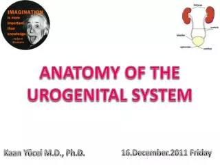
ANATOMY OF THE UROGENITAL SYSTEM. Kaan Yücel M.D., Ph.D. 16.December.2011 Friday. KIDNEYS. R emove excess water, salts, and wastes of protein metabolism from the blood R eturn nutrients and chemicals to the blood. L ie retroperitoneally on the posterior abdominal wall,

E N D
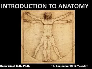
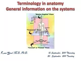
ANATOMY OF THE UROGENITAL SYSTEM • Kaan Yücel M.D., Ph.D. • 16.December.2011 Friday
KIDNEYS Removeexcess water, salts, and wastes of protein metabolism from the blood Returnnutrients and chemicals to the blood. Lieretroperitoneally on the posterior abdominal wall, one on each side of the vertebral column.
At the concave medial margin of each kidney is a vertical cleft, the renal hilum. The renal hilum is the entrance to a space within the kidney, the renal sinus. Structures that serve the kidneys (vessels, nerves, and structures that drain urine from the kidney) enter and exit the renal sinus through the renal hilum.
Each kidney has anterior and posterior surfaces, medial and lateral margins, and superior and inferior poles. The renal pelvis is the flattened, funnel-shaped expansion of the superior end of the ureter.
The renal pelvis receives two or three major calices (calyces), each of which divides into two or three minor calices. Each minor calyx is indented by a renal papilla, the apex of the renal pyramid, from which the urine is excreted. The pyramids and their associated cortex form the lobes of the kidney.
SUPRARENAL GLANDS Locatedbetween the superomedial aspects of the kidneys & diaphragm, where they are surrounded by connective tissue containing considerable perinephric fat.
Suprarenalcortex secretes corticosteroids and androgens. These hormones cause the kidneys to retain sodium and water in response to stress, increasing the blood volume and blood pressure. They also affect muscles and organs such as the heart and lungs.
Suprarenalmedullais a mass of nervous tissue associated with the sympathetic nervous system. The chromaffin cells of the medulla secrete catecholamines (mostly epinephrine) into the bloodstream in response to signals from presynaptic neurons. The powerful medullary hormones epinephrine (adrenaline) and norepinephrine (noradrenaline) activate the body to a flight-or-fight status in response to traumatic stress. They also increase heart rate and blood pressure, dilate the bronchioles, and change blood flow patterns, preparing for physical exertion.
PELVIC VISCERA Pelvicviscera include the distal parts of the urinary system and digestive tract, and the reproductive system. The bladder and rectum—true pelvic viscera—are inferior continuations of systems encountered in the abdomen.
Except for features related to sharing of the male urethra by the urinary and reproductive tracts, and physical relationships to the respective reproductive organs, there are relatively few distinctions between the male and female pelvic urinary and digestive organs.
Urinary Organs • Pelvic urinary organs: • Pelvic portions of the ureters, which carry urine from the kidneys. • Urinary bladder, which temporarily stores urine. • Urethra, which conducts urine from the bladder to the exterior.
URETERS Muscularducts (25-30 cm long) with narrow lumina that carry urine from the kidneys to the urinary bladder. Run inferiorly from apices of the renal pelves at the hila of the kidneys. Along the lateral wall of the pelvis and enter the urinary bladder.
URINARY BLADDER Ahollow viscus with strong muscular walls Characterizedby its distensibility Temporaryreservoir for urine Variesin size, shape, position, and relationships according to its content and the state of neighboring viscera When empty, the adult urinary bladder is lies on the pubic bones and pubic symphysis anteriorly and the prostate (males) or anterior wall of the vagina posteriorly.
Toward the neck of the male bladder, the muscle fibers form the involuntary internal urethral sphincter. This sphincter contracts during ejaculation to prevent retrograde ejaculation (ejaculatory reflux) of semen into the bladder.
MALE URETHRA Musculartube that conveys urine from internal urethral orifice of the urinary bladder to external urethral orifice, located at the tip of the glans penis in males. Alsoprovides an exit for semen (sperms and glandular secretions).
FEMALE URETHRA Passes anteroinferiorly from the internal urethral orifice of the urinary bladder, posterior and then inferior to the pubic symphysis, to the external urethral orifice.
The musculature surrounding the internal urethral orifice of the female bladder is not organized into an internal sphincter. In females, the external urethral orifice is located in the vestibule, the cleft between the labia minora of the external genitalia, directly anterior to the vaginal orifice. The urethra lies anterior to the vagina (forming an elevation in the anterior vaginal wall).
MALE GENITAL SYSTEM • The reproductive system in men has components in the abdomen, pelvis, and perineum. • The major components are a testis, epididymis, ductus deferens, and ejaculatory duct on each side, and the urethra and penis in the midline. • In addition, three types of accessory glands are associated with the system: • a single prostate; • a pair of seminal vesicles; and • a pair of bulbourethral glands.
SEMINAL GLANDS • Each seminal gland (vesicle) is an elongated structure that lies between the fundus of the bladder and the rectum. • The seminal glands are obliquely placed superior to the prostate and do not store sperms, as the “vesicle” term implies. • They secrete a thick alkaline fluid with fructose (an energy source for sperms) and a coagulating agent that mixes with the sperms as they pass into the ejaculatory ducts and urethra.
EJACULATORY DUCTS Slender tubes Unionof ducts of the seminal glands with ductusdeferentes
PROSTATE Largestaccessory gland of the male reproductive system Firm, walnutsizep Surroundsthe prostatic urethra. The glandular part makes up approximately two thirds of the prostate; the other third is fibromuscular.
BULBOURETHRAL GLANDS • Lieposterolateral to the intermediate part of the urethra • Their ducts open into the proximal part of the penis
SPERMATIC CORD • Containsstructures running to and from the testis • Passes through the inguinal canal • Endsin the scrotum at the posterior border of the testis
SCROTUM Acutaneous sac consisting of 2 layers: heavily pigmented skin and the closely related dartos fascia, a fat-free fascial layer including smooth muscle fibers (dartos muscle) responsible for the rugose (wrinkled) appearance of the scrotum. Because the dartos muscle attaches to the skin, its contraction causes the scrotum to wrinkle when cold, and assisting the muscles in holding the testes closer to the body, all of which reduces heat loss. The scrotum is the male homologue of the labia majora in women.
TESTIS (TESTICLES) Male gonads—paired ovoid reproductive glands that produce sperms (spermatozoa) and male hormones, primarily testosterone. Originallydevelop high on the posterior abdominal wall and then descend, normally before birth, through the inguinal canal in the anterior abdominal wall and into the scrotum of the perineum.
EPIDIDYMIS An elongated structure on the posterior surface of the testis. Efferent ductules of the testis transport newly developed sperms to the epididymis. At the tail of the epididymis, the ductus deferens begins as the continuation of the epididymal duct.
PENIS • Suspendedfrom the front and sides of the pubic arch and containing the greater part of the urethra. • Consistsof a root and body. • Bodyof penisis entirely covered by skin; the tip of the body is covered by the glans penis. • The external urethral orifice is a sagittal slit, normally positioned at the tip of the glans.
FEMALE GENITAL SYSTEM The female internal genital organs include the ovaries, uterine tubes, uterus, and vagina.
OVARIES • Almond-shaped and -sized female gonads in which the oocytes develop.Lie adjacent to the lateral pelvic wall. • Alsoendocrine glands that produce reproductive hormones. • Like the testes in men, the ovaries develop high on the posterior abdominal wall and then descend before birth, bringing with them their vessels, lymphatics, and nerves. • Unlike the testes, the ovaries do not migrate through the inguinal canal into the perineum, but stop short and assume a position on the lateral wall of the pelvic cavity.
UTERINE TUBES • Conductthe oocyte, discharged monthly from an ovary during child-bearing years, from the periovarian peritoneal cavity to the uterine cavity. • Providethe usual site of fertilization. • Extendlaterally from the uterine horns and open into the peritoneal cavity near the ovaries.
The uterine tubes are divisible into four parts, from lateral to medial: Infundibulum: opens into the peritoneal cavity through the abdominal ostium. Ampulla: widest and longest part of the tube, which begins at the medial end of the infundibulum; fertilization of the oocyte usually occurs in the ampulla. Isthmus: t thick-walled part of the tube, which enters the uterine horn. Uterine part: short intramural segment, opens via the uterine ostium into uterine cavity at the uterine horn.
UTERUS Theuterus(womb) is a thick-walled, pear-shaped, hollow muscular organ. The embryo and fetus develop in the uterus. Its muscular walls adapt to the growth of the fetus and then provide the power for its expulsion during childbirth. The non-gravid (non-pregnant) uterus usually lies in the lesser pelvis, with its body lying on the urinary bladder and its cervix between the urinary bladder and rectum.
The uterus is a very dynamic structure, the size and proportions of which change during the various changes of life. • When the bladder is empty, the uterus typically lies in a nearly transverse plane. • The position of the uterus changes with the degree of fullness of the bladder and rectum, and stage of pregnancy.
The uterus is divisible into two main parts: the body and cervix. • The uterine horns (L. cornua) are the superolateral regions of the uterine cavity, where the uterine tubes enter. • The wall of the body of the uterus consists of three coats, or layers: • Perimetrium • Myometrium • Endometrium
VAGINA Adistensible musculomembranous tube, extends from the middle cervix of the uterus to the vaginal orifice, the opening at the inferior end of the vagina.
The vagina lies posterior to the urinary bladder and urethra, the latter projecting into its inferior anterior wall. • It lies anterior to the rectum.
EXTERNAL PART OF THE FEMALE GENITAL SYSTEM • In women, the clitorisand vestibular apparatus, together with a number of skin and tissue folds, form the vulva. • On either side of the midline are two thin folds of skin termed the labia minora.
Lateral to the labia minora are two broad folds, the labia majora, which unite anteriorly to form the mons pubis. • The mons pubis overlies the inferior aspect of the pubic symphysis and is anterior to the vestibule and the clitoris.