Download
1 / 41
420 likes | 669 Vues
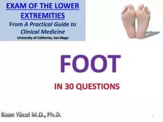
Exam of the Lower Extremities From A Practical Guide to Clinical Medicine University of California, San Diego. FOOT. IN 30 QUESTIONS. Kaan Yücel M.D., Ph.D. INTRODUCTION TO FOOT. R egion of the lower limb distal to ankle joint S ubdivided into Ankle Metatarsus D igits

E N D
Exam of the Lower Extremities From A Practical Guide to Clinical Medicine University of California, San Diego • FOOT • IN 30 QUESTIONS • Kaan Yücel M.D., Ph.D.
INTRODUCTION TO FOOT Region of the lower limb distaltoankle joint Subdivided into Ankle Metatarsus Digits Superior surface (dorsum of foot) Inferior surface (sole)
INTRODUCTION TO FOOT • Body's point of contact with the ground • Provides a stable platform for upright stance. • Supports the body weight and provides leverage for walking and running. • Unique -constructed in the form of arches, adapt its shape to unevensurfaces. • Serves as a resilient spring to absorb shocks, such as in jumping.
SKIN & SUBCUTANEOUS TISSUE • IN 10 QUESTIONS
1…skin of the foot Skin of the dorsum of the foot thinner and less sensitive than that of the sole The subcutaneous tissue is loose deep to the dorsal skin Edema most marked on the dorsal skin especially anterior to and around the medial malleolus Entire sole is sensitive (“ticklish”), especially the thinner-skinned area underlying the arch of the foot. Neurosci Res. 2010 Dec;68(4):285-9. Neurogenesis in the dentate gyrus of the rat hippocampus enhanced by tickling stimulation with positive emotion. Ticklish? gargalesis
2. Deep fascia of the foot & Plantar fascia Thin where it is continuous proximally with the inferior extensor retinaculum. Over the lateral and posterior aspects of the footcontinuous with the plantar fascia, the deep fascia of the sole. Plantar fascia holds the parts of the foot together, helps protect the sole from injury, and helps support the longitudinal arches of the foot.
3. Compartments by the deep fascia Midfoot and forefoot Medial compartment of the sole Central compartment of the sole Lateral compartment of the sole Forefoot 4th compartment interosseous compartment of the foot Dorsal compartment of the foot 5th compartment between dorsal fascia of the foot & tarsal bones & dorsal interosseous fascia of the midfoot and forefoot http://home.comcast.net/~wnor/soleoffoot.htm Transverse section of the foot: 1, lateral compartment; 2, central compartment; 3, medial compartment; 4, interosseous compartment. Arrows indicate the high-pressure areas that often lead to foot ulceration.
4. Tarsal tunnel • Formed on the posteromedial side of the ankle by: • A depression formed by • Medial malleolus of tibia Medial &posterior surfaces of talus • Inferior surface of sustentaculum tali Medial surface of calcaneus • Overlying flexor retinaculum • Contents from ant. to post. • (Tom Dick ANdHarry) • Tibialis posterior tendon • Flexor digitorumlongus tendon • Posterior tibial artery • Tibial nerve • Flexor hallucislongus tendon
5. Flexor retinaculum Attaches above to medial malleolus below to calcaneus 2 compartments on the posterior surface of medial malleolus for tibialis posterior& flexor digitorum longus tendons Laterally posterior tibial artery ,veins& tibial nerve through tarsal tunnel into sole of foot Lateral to tibial nerve tendon of flexor hallucis longus Pulse of posterior tibial artery through flexor retinaculum midway between medial malleolus and calcaneus
6. Extensor retinacula Prevent tendon bowing during extension of the foot and toes Superior extensor retinaculum Superior to the ankle Attached to anterior borders of the fibula & tibia Inferior retinaculum Y-shaped Attached to lateral side of calcaneus Extensor digitorum longus & fibularis tertius tendons Medially Dorsalis pedis artery Extensor hallucis longus tendon tibialis anterior tendon pass under extensor retinacula
7. Fibular (Peroneal) retinacula Bind the tendons of fibularis longus &fibularis brevis to lateral side of the foot Superior fibular retinaculum Between lateral malleolus & calcaneus Inferior fibular retinaculum Attaches to lateral surface of calcaneus around fibular trochlea
8. Plantar aponeurosis A thickening of deep fascia in the sole of thefoot A thick centralpart Weaker medial and lateralparts Tougher, denser, andelongatedthanpalmaraponeurosis Anchored to the medial process of calcanealtuberosity Supports the longitudinal arch of the foot & protects deeper structures in the sole
9. Fibrous sheaths of toes Flexor digitorum longus, flexor digitorum brevis, & flexor hallucis longustendons enter fibrous digital sheaths on plantar aspect of digits. Formed by fibrous arches & cruciate (cross-shaped) ligaments Hold the tendons to the bony plane and prevent tendon bowing when the toes are flexed.
10. Extensor hoods Extensor digitorum longus, extensor digitorum brevis, and extensor hallucis longusexpand over the proximal phalanges to form complex dorsal digital expansions ("extensor hoods"). Manyof the intrinsic muscles of the foot insert here. Thisallows the forces from these muscles to be distributed over the toes to cause flexion of the metatarsophalangeal joints while at the same time extending the interphalangeal joints.
MUSCLES OF THE FOOT • IN 4 QUESTIONS
20 individual muscles of thefoot • 14 located on the plantaraspect • 2 on the dorsalaspect • 4 intermediate in position • Plantar muscles function primarily as a group during the support phase of stance, maintaining the arches of the foot • Few delicate functionscomparedtohandmuscles • Concerned with supporting the arches of thefoot
Muscles in the dorsum Extensor digitorum brevis Attached to a roughened area on the superolateral surface of the calcaneus lateral to the tarsal sinus Extensor hallucis brevis The part of the muscle associated with the great toe Deepfibularnerve
Muscles in the sole Organized into four layers From superficial to deep or plantar to dorsal First layer Medial to lateral Abductor hallucis Flexor digitorum brevisAbductor digiti minimi Second layer Quadratus plantae Lumbrical muscles Third layer Flexor hallucis brevis Adductor hallucis Flexor digiti minimi brevis Fourth layer Dorsal & plantar interossei
Functions & innervations of the muscles of the foot 1st layer Medialplantarnerve (S2, S3) Abductorhallucis Flexordigitorumbrevis Abductordigiti minimi Lateralplantarnerve (S2, S3) 2nd layer Assistsflexordigitorumlongus in flexinglateralfourdigits Quadratusplantae Flexproximalphalanges, extendmiddle& distalphalanges of lateralfourdigits Lumbricals 3rdlayer 4th layer Plantarinterossei (threemuscles) Adductdigits (2-4) andflexmetatarsophalangealjoints Dorsalinterossei (fourmuscles) Abductdigits (2-4) andflexmetatarsophalangealjoints Flexorhallucisbrevis Adductorhallucis deepbranch Flexordigit minimi brevissuperficialbranch
ARTERIES OF THE FOOT • IN 5 QUESTIONS
Supply of the foot Terminal branches of anterior tibial artery &posterior tibial artery dorsalis pedis artery&plantar arteries Posterior tibial artery in the sole divides into lateral & medial plantar arteries Lateral plantar artery +Dorsalis pedis artery Deep plantar arch
Lateral & medial plantar arteries • Lateral plantar artery • Major branches of the deep plantar arch • Digital branch tolateralside of little toe • 4 plantar metatarsal arteries • 3 perforating arteries • Medial plantar artery • Passes deep to the abductor hallucis • Ends by joining the digital branch of the deep plantar arch • Superficial branch
Dorsalis pedis artery Major source of blood supply toforefoot Continuation of anteriortibialartery Begins midway betweenmalleoli Runsdeeptoinferior extensor retinaculum betweenextensor hallucis longus& extensor digitorum longus tendons on the dorsum of thefoot
Dorsalis pedis artery • Passesinferiorly as • deepplantarartery • Joinsdeep plantar arch in the sole of the foot. • Branches • lateral and medial tarsalbranches • arcuateartery • first dorsal metatarsalartery
VEINS OF THE FOOT • IN 2 QUESTIONS
As in the rest of the lower limb, there are both superficial and deep veins in the foot. Unlike the leg and thigh, however, the venous drainage of the foot is primarily to the major superficialveins
Dorsalmetatarsalveins Dorsaldigitalveins Dorsalvenous network of thefoot Dorsalvenousarch of thefoot Medialside Lateralside Great saphenousvein Small saphenousvein
NERVES OF THE FOOT • IN 8 QUESTIONS
Tibial nerve • Deep fibular nerve • Superficial fibular nerve • Sural nerve • Saphenous nerve • Tibial nerve • All intrinsic muscles of the foot except for the extensor digitorum brevis, innervated by the deep fibular nerve • Deep fibular nerve often also contributes to innervation of the first and second dorsal interossei.
Tibial nerve Enters the foot through tarsal tunnel posterior to medial malleolus Lateral to posterior tibial artery Medial calcaneal branchesheel Midway between medial malleolus & heel large Medial plantar nerve smaller Lateral plantar nerve
Medialplantar nerves Larger & moreanterior terminal branchArisesdeeptotheflexorretinaculum Majorsensorynerve in thesole Comparedtolateralplantarnerve, supplies more skin area but fewer muscles. Its distribution to both skin and muscles of comparable to that of the median nerve in the hand. Skin on most of theanteriortwo-thirds of the sole andadjacentsurfaces of themedialthreeandone-halftoes, whichincludesthegreattoe. Fourintrinsicmuscles- abductorhallucis, flexordigitorumbrevis, flexorhallucisbrevis, andthefirstlumbrical.
Lateral plantar nerve Smaller & more posterior terminal branch Also courses deep to the abductor hallucis but runs anterolaterally between the 1st and 2nd layers of plantar muscles, on the medial side of the lateral plantar artery. Ends @ lateral compartment, dividing into superficial and deep branches.
Lateral plantar nerve Superficialbranch Twoplantardigitalnerves (onecommonandoneproper) Skin of theplantaraspects of thelateraloneand a halfdigits, thedorsal skin and nail beds of theirdistalphalanges, and skin of the sole proximaltothem.
Deep fibular nerve Passes deep toextensorretinaculum Entersdorsal aspect of the foot on the lateral side of dorsalis pedis artery. Muscles on dorsum of thefoot Whenfinally emerges as a cutaneous nerve, so far distal in the foot ; small area of skin remains available for innervation: web of skin between and contiguous sides of the 1st and 2nd toes Divides into two dorsal digitalnerves
Superficial fibular nerve Emerges as a cutaneous nerve about two thirds of the way down the leg Skin on the anterolateral aspect of leg Divides into Medial and intermediate dorsal cutaneous nerves Most of the skin on the dorsum of the foot Terminal branches Dorsal digital nerves (common & proper) Skin of the proximal aspect of the medial half of the great toe & that of the lateral three and a half digits.
Sural nerve • Cutaneous branch of tibial nerve Originates high in the leg • Sural nerve formation: • Medial sural cutaneous nerve (from the tibial nerve) • Sural communicating branch of common fibular nerve • Accompanies small saphenous vein Enters the foot posterior to the lateral malleolus • ankle joint &skin on the lateral side of the foot and dorsolateral surface of the little toe.
Saphenous nerve • Longest & most widely distributed cutaneous branch of the femoral nerve • Only branch to extend beyond the knee • Originates in the thigh • Skin and fascia on the anteromedial aspect of the leg,ankle joint • Skin along the medial side of the foot as far anteriorly as the head of the 1st metatarsal , medial side of the proximal foot
Cutaneous innervation of the foot Medially Saphenous nervedistally to the head of 1st metatarsal Superiorly (dorsum of foot) Superficial (primarily) Deep fibular nerves Inferiorly (sole of foot) Medial & lateral plantar nerves Laterally Sural nerve Posteriorly (heel) Medial & lateral calcaneal branches of tibial & sural nerves, respectively
LYMPHATIC DRAINAGE OF THE FOOT
Medialsuperficial lymphaticvessels medialside of the dorsum& sole of thefoot Accompanygreatsaphenous vein Lateralsuperficial lymphatic vessels lateralside of the dorsum& sole of thefoot Accompanysmallsaphenous vein Deepinguinal lymphnodes Iliaclymphnodes