Download
1 / 19
210 likes | 945 Vues
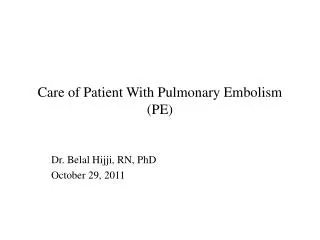
Care of Patient With Pulmonary Embolism (PE). Dr. Belal Hijji, RN, PhD October 29, 2011. Learning Outcomes. At the end of this lecture, students will be able to: Describe PE, its pathophysiological changes, and discuss its clinical manifestations.

E N D
Care of Patient With Pulmonary Embolism (PE) Dr. Belal Hijji, RN, PhD October 29, 2011
Learning Outcomes At the end of this lecture, students will be able to: • Describe PE, its pathophysiological changes, and discuss its clinical manifestations. • Identify the diagnostic test that may be used to diagnose PE. • Discuss the medical and nursing management of PE.
Introduction • Pulmonary embolism (PE) is an obstruction of the pulmonary artery (next slide) or one of its branches by a thrombus (or thrombi) that originates somewhere in the venous system. • The types of emboli could be a blood clot (most common), air, fat, amniotic, fluid, and septic (from bacterial invasion of the thrombus). • PE is often associated with trauma, surgery (orthopedic), pregnancy, heart failure, age > 50 years, hypercoagulable states, and prolonged immobility. • Most thrombi originate in the deep veins of the legs; other sites include the pelvic veins and the heart’s right atrium. • An enlarged right atrium in fibrillation causes blood to stagnate[ركود] and form clots that may travel into the pulmonary circulation causing PE.
Pathophysiology • When there is a complete or partial obstruction of a pulmonary artery or its branches by a thrombus, the alveolar dead space (next slide) is increased. The area, although continuing to be ventilated, receives little or no blood flow, resulting in impaired or absent gas exchange. • In addition, various substances are released from the clot and surrounding area, causing regional blood vessels and bronchioles to constrict. This causes an increase in pulmonary vascular resistance. This results in an increase in pulmonary arterial pressure and, in turn, an increase in right ventricular work to maintain pulmonary blood flow. • When the work requirements of the right ventricle exceed its capacity, right ventricular failure occurs, leading to a decrease in cardiac output followed by a decrease in systemic blood pressure and the development of shock.
Alveolar dead space: A well-ventilated part of the lung is not receiving blood flow. The air reaching that region of the lung is therefore wasted since it cannot participate in gas exchange, thus the alveoli are considered dead.
Clinical Manifestations • The symptoms of PE depend on the size of the thrombus and the area of the pulmonary artery occluded by the thrombus. • Dyspnea is the most frequent symptom; while tachypnea is the most frequent sign. Chest pain is common and is usually sudden and pleuritic. Other symptoms include anxiety, fever, tachycardia, apprehension, cough, diaphoresis, hemoptysis, and syncope. • Deep venous thrombosis is closely associated with the development of PE. Typically, patients report sudden onset of pain and/or swelling and warmth of the proximal or distal extremity, skin discoloration, and superficial vein distention.
Assessment and Diagnostic Findings • Early recognition and diagnosis of PE are priorities as death commonly occurs within 1 hour of symptoms. • The diagnostic workup includes a ventilation–perfusion scan, pulmonary angiography, chest x-ray (may show infiltrates, atelectasis, elevation of the diaphragm on the affected side, or a pleural effusion), ECG (sinus tachycardia, PRinterval depression, and nonspecific T-wave changes), and arterial blood gas analysis (may show hypoxemia and hypocapnia (from tachypnea)). • If lung scan results are not definitive, pulmonary angiography is the gold standard for the diagnosis of PE.
Medical Management • Emergency management. • Nasal oxygen to relieve hypoxemia, respiratory distress, and central cyanosis. • Intravenous infusion lines to administer medications or fluids. • A perfusion scan, arterial blood gas determinations are performed. Pulmonary angiography may be performed. • Hypotension is treated by a slow infusion of dobutamine (Dobutrex). • The ECG is monitored continuously for dysrhythmias which may occur suddenly. • Digitalis glycosides, intravenous diuretics, and antiarrhythmic agents may be indicated. • Blood is drawn for serum electrolytes and complete blood count. • Intubation and mechanical ventilation may be performed based on clinical assessment and arterial blood gas analysis.
Emergency management (Continued…). • In case of hypotension, a Foley’s catheter is inserted to monitor urinary output. • Small doses of intravenous morphine to relieve the patient’s anxiety, to alleviate chest discomfort, to improve tolerance of the endotracheal tube, and to ease adaptation to the mechanical ventilator. • Pharmacologic therapy. (Anticoagulation) • Heparin is used to prevent recurrence of emboli. The dose is an intravenous bolus of 5,000 to 10,000 units, followed by a continuous infusion at a rate of 18 U/kg per hour. The rate is reduced in patients with a high risk of bleeding. Heparin is usually administered for 5 to 7 days. • Warfarin sodium administration is begun within 24 hours after initiation of heparin therapy because its onset of action is 4 to 5 days. Warfarin is usually continued for 3 to 6 months. Anticoagulation therapy is contraindicated in patients who are at risk for bleeding (eg, those with gastrointestinal conditions or with postoperative or postpartum bleeding).
Pharmacologic therapy (Continued…). Thrombolytic therapy • Streptokinase may be used in patients who are hypotensive and have significant hypoxemia. It resolves the thrombi or emboli more quickly and reduces pulmonary hypertension and improves perfusion, oxygenation, and cardiac output. • Is initiated after stopping heparin. During therapy, all but essential invasive procedures are avoided because of potential bleeding. • Cessation necessitates the initiation of anticoagulants.
Nursing Management • Minimizing The Risk of Pulmonary Embolism • A major responsibility of the nurse is to identify patients at high risk for PE and to minimize the risk of PE in all patients. Therefore, the nurse must give attention to conditions predisposing to a slowing of venous return (i.e. prolonged immobilization, prolonged periods of sitting/traveling, varicose veins, spinal cord injury), hypercoagulability due to release of tissue thromboplastin after injury/surgery (i.e. pancreatic, GI, GU, breast, or lung tumor, increased platelet count in polycythemia), venous endothelial disease (i.e. thrombophlebitis, foreign bodies such as IV/central venous catheters)
Preventing Thrombus Formation. The nurse: • encourages ambulation and active and passive leg exercises to prevent venous stasis in patients on bed rest and to help increase venous flow. • discourages the patient against sitting or lying in bed for prolonged periods, crossing the legs, and wearing constricting clothing. Legs • discourages legs dangling or feet placed in a dependent position while sitting on the edge of the bed; instead, the patient’s feet should rest on a chair. • Should not leave intravenous catheters in place for prolonged periods.
Assessing Potential For Pulmonary Embolism. The nurse should: • examine patients who are at risk for developing PE for a positive Homans’ sign (pain in the calf as the foot is sharply dorsiflexed), which may or may not indicate impending thrombosis of the leg veins. A positive Homans’ sign may indicate DVT.
Monitoring Thrombolytic Therapy. The nurse: • keeps the patient on bed rest • assesses vital signs Q2H. • ensures that tests to determine prothrombin time or partial thromboplastin time are performed 3 to 4 hours after the thrombolytic infusion is started to confirm that the fibrinolytic systems have been activated. • ensures that only essential venipunctures are performed because of the prolonged clotting time, and manual pressure is applied to any puncture site for at least 30 minutes. • uses pulse oximetry to monitor changes in oxygenation. • immediately discontinues the infusion if uncontrolled bleeding occurs.
Managing Chest Pain. The nurse: • Places the patient in a semi-Fowler’s position which is more comfortable for breathing. • continues to turn the patient frequently and repositioning him to improve the ventilation–perfusion ratio in the lung. • Administers opioid analgesics as prescribed for pain.
Managing Oxygen Therapy. The nurse: • gives careful attention the proper use of oxygen and ensures that the patient understands the need for continuous oxygen therapy. • assesses the patient frequently for signs of hypoxemia and monitors the pulse oximetry values to evaluate the effectiveness of the oxygen therapy. • encourages deep breathing and performs incentive spirometry to minimize or prevent atelectasis and improve ventilation.
Managing Anxiety. The nurse: • encourages the stabilized patient to talk about any fears or concerns related to this frightening episode. • answers the patient’s and family’s questions concisely and accurately. • explains the therapy, and describes how to recognize untoward effects early.