Download
1 / 39
390 likes | 409 Vues
This article provides an overview of the upper respiratory tract, including the nasal passages, paranasal sinuses, and pharynx, as well as the lower respiratory tract, including the trachea and lungs. It also discusses respiration, respiration rates, asthma, exercise-induced bronchospasm, pleurisy, and upper respiratory infections.

E N D
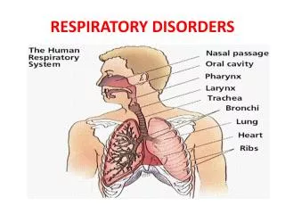
Upper Respiratory Tract • Nasal passages • Paranasal sinuses • Pharynx • Nasopharynx • Oropharynx • Larynx • Function • Warming • Humidifying • Filtering air
Lower Respiratory Tract • Trachea • Lungs • Right = 3 lobes • Left = 2 lobes • Bronchi • Bronchioles • Terminal alveoli
Respiration (simultaneous) • Ventilation • Air moves through respiratory tract • Oxygenation • Exchange of gases in the alveolar-capillary beds
Inspiration • Diaphragm and intercostal muscles contract • Thoracic cavity expands = negative pressure = movement of air into lungs • Alveolar pressure= atmospheric pressure = intercostal stretch receptors fire • Inspiration ceases
Expiration • Elastic recoil of thoracic cage = passive expiration
Respiration Rates • Unlabored = 12 to 20 /minute • Newborn – 30 to 60 • Infant – 25 to 40 • Toddler – 20 to 30 • Young children – 20 to 25 • Older children – 15 to 20 • Dyspnea = difficulty breathing • Apnea = absence • Obstructive sleep apnea • Tachypnea = >24 /minute “rapid breathing” • Pulmonary embolism • Enlarged liver or spleen
Respiration Rates • Hyperpnea = tachypnea with large breaths = hyperventilation • Anxiety • Diabetic ketoacidosis (DKA) • Bradypnea = <12 /minute = “slow breathing” • Well conditioned athletes • Electrolyte imbalance • Hypopnea = “shallow, slow breaths” • Rib fracture
Asthma • Chronic inflammatory disease • Intermittent acute bronchospasms • Affects 25 million people in the US, 300 million worldwide • Causes epithelial cell damage and fibrous changes in bronchioles = reduction in airflow • Sighs and symptoms • Dyspnea (difficulty breathing) • Coughing • Wheezing • Chest tightness • Prolonged expiration • Panting speech • Posture – leaning forward to assist breathing • Cyanosis (blue/gray skin coloring) • Seizure – severe cases
Asthma • Causes • Allergens • Respiratory infections • Cold/dry air • Drugs • Emotional states • Treatment • Long-acting albuterol inhaler (often combined with corticosteroids) • Short-acting albuterol inhaler • Policies and Procedures • Have a written action plan for each patient with asthma
Asthma • Action plan should include: • Physician’s name and contact information • Long-term and quick relief medications (names and doses) • Personal best peak expiratory flow rate (PEFR) • Physician’s instructions for managing patient’s acute asthma attack • Rescue inhaler (number of puffs/dose, time between doses, number of doses) • Nebulizer • Supplemental oxygen • Criteria for seeking EMS • RTP criteria (return to play)
Asthma • Managing an acute asthma attack • Patient in seated position • Instruct patient to take deep breaths • Exhale through pursed lips (increases pressure throughout airway) • Attempt to calm emotionally • Take PEFR (peak exp. flow rate) • Administer short-acting ß2 agonists (inhaler) • If prescribed and indicated • Takes 5 to 15 minutes to act • Takes up to 3 doses in a 1 hour period • Take PEFR again • Recovery should occur gradually in about 1 hour • Pulse oximeter can monitor oxygen saturation • Administer supplemental oxygen SpO2 < 92% • Treat as emergency if little to no recovery
Asthma • Severe attack may cause: • Pneumothorax (collapsed lung) • Acute right heart failure • Hypoxemia (low O2 level in blood) • Metabolic collapse (muscles cannot function) • Signs and symptoms • Use of accessory muscles to breath • Cyanosis • Confusion • Sweating • Poor air movement
Exercise-Induced Bronchospasm • AKA exercise-induced asthma • Affects 15% of population • Occurs 5 to 10 minutes into exercise • Becomes progressively worse with activity • Spontaneous recovery occurs 30 to 60 of rest after ceasing activity • Does not produce chronic inflammation in the bronchioles • Sign and symptoms • Unusual dyspnea (difficulty breathing) • Central chest pain or tightness during exercise • Coughing after strenuous exercise
Exercise-Induced Bronchospasm • Factors that exacerbate • Allergens • Infection • Pollution • Cool or dry air mouth breathing • Prevention • Preventive medication • Environmental precautions • Prolonged warm up (60 minutes) • 10 to 15 minutes at 50%, MHR • Rest 15 minutes prior to activity • Treatment • Same as an asthma attack
Pleurisy • AKA pleuritis or pleuritic pain • Any inflammation of the pleura that causes pain
Upper Respiratory Infections • Common signs and symptoms • Rhinitis (nose inflammation) • Rhinorrhea (runny nose) • Sore throat • Nonproductive cough • Sneeze • Headache • Malaise (feeling run down, low energy) • Chills • Low grade fever • Laryngitis (inflammation of the larynx) • Duration • 7 to 10 days
Upper Respiratory Infections • Complications • Ear infections • Sinus infections • May include tooth pain • Facial or sinus pressure and pain • Nasal congestion or discharge • Foul breath • Virus • Contagious • Active for at least 8 days following initial infection • Communicability highest in first 72 hours • Prevention • Avoid contact with persons exhibiting sx/sy of URI • Frequent hand washing • 20 seconds or sing the “happy birthday” song
Upper Respiratory Infections • Treatment • Rest • Fluids • Nutrition • OTC’s • Caution patients from “stacking” meds • Do not use aspirin with anyone 18 and younger • Reye’s syndrome = altered mental state and severe vomiting • “neck rule” (symptoms above neck = ok to exercise) • Hold an additional 24 hours after sx/sy have ceased • Physician referral after 10 days • Antibiotics only work if bacterial
Acute Bronchitis • Lower respiratory infection • Viral most common • Typically preceded by URI • Productive cough • Wheezing • Treatment • OTC’s • Rest • Hydration • Possibly short-acting inhaler • RTP • “neck rule” • Gradual return
Pneumonia • Lower respiratory infection • Bacterial, viral or fungal • Signs and symptoms • Productive cough • Dyspnea • Pleuritic chest pain • Fever • Myalgia • Malaise • Tachypnea • Tachycardia • Crackles with auscultation • Dullness with percussion
Pneumonia • Treatment • Refer to physician • OTC’s • Antibiotics • Rest • Fluids • Supplemental oxygen • RTP • Similar to that of acute bronchitis, however, would be longer
Influenza • AKA flu • Occur in fall, peaking in the winter, lasting into spring • Three recognized strains – A, B, and C • A = most commonly associated with large outbreaks • Transmitted • Person to person through respiratory secretions • Incubation period of 2 days • Treatment • Antiviral – occasionally (administered at first sign of symptoms) • OTC’s • RTP • “neck rule” • Gradual increase of intensity and duration
Tuberculosis • Pulmonary tuberculosis (TB) • Highly contagious • Develops granular tumors in the infected tissue • May be dormant for years before causing active disease • Suspend from activity and refer to a physician
Tuberculosis • Signs and symptoms • Fatigue • Fever >100° • Chills • Night sweats • Weight loss • Cough • Hemoptysis • Chest pain • SOB • Wheezing • Crackles or wheezing with auscultation • Enlarged and tender lymph nodes • Clubbing of fingers and toes • Bold = key symptoms
Chronic Obstructive Pulmonary Disease (COPD) • Examples • Chronic bronchitis • Hypoxemia • Cyanosis • Productive cough (greater in mornings and evenings) • Cough > 3 months for 2 consecutive years with no other explanation • Emphysema • Complication of chronic pulmonary disease and smoking • Damage is irreversible and prognosis is poor • Cystic fibrosis • Inherited • Damage to lungs and digestive system • No cure • Some may live into 40’s or 50’s, commonly to 20’s and 30’s
Atelectasis • Not a disease but a complication • Collapse of a lung segment’s alveoli • Common – more than 200,000 cases per year • RTP – depends on amount of damage
Drowning/near drowning • Inhaled water physically damages lung tissue • Leads to atelectasis • Reflex spasm of larynx – die from asphyxia • Time of immersion and temperature of water determines extent of damage for a near drowning victim • Neurological complications • Renal complications • Rapid, progressive respiratory failure can occur 12 to 24 hours after rescue • Always perform life saving strategies • Always transport
Pneumothorax • Pneumothorax AKA “collapsed lung • Air in the pleural space • Negative pressure that holds lungs inflated is lost • Lung retracts toward bronchial tree • Symptoms and symptoms • Decreased or absent breath sounds • Acute pleuritic chest pain • Dyspnea • Hyperpnea • Crepitus
Pneumothorax • Management • Splint by hugging a pillow • Calm the patient to control coughing or gasping for air • Monitor vital signs • Seal any open wounds • Transport • RTP • May occur within days of discharge • If other injuries permit
Hemothorax • Blood enters the pleural cavity • Splashing may be heard on auscultation in the lower lobes with changes in posture • Blood and air = hemopneumothorax
Flail Chest Injury • Results from multiple anterior or posterior rib fractures • Affected region collapses on inspiration • Affected region bulges on expiration • Medical emergency • Often accompanied by pneumothorax
Pediatric Concerns • Asthma • Cystic fibrosis • Neuromuscular diseases • May affect respiratory muscles and neural control of breathing • Scoliosis • May impair inflation of one or both lungs • Pertussis “whooping cough” • Highly contagious • Asymptomatic incubation period (7 to 21 days) • Catarrhal stage – 1 to 2 weeks, resembles a simple URI • Most contagious period • Paroxysmal state • Whooping cough appears and increases over a week • 6 to 10 weeks • Convalescent stage • Gradual cessation of cough over 2 to 3 weeks
Therapeutic Interventions • Supplemental Oxygen (possible sickle cell distress) • Assemble tank and regulator • Hand-tighten the regulator • Check tank pressure – turn valve stem one complete turn • 500 psi = 25% full = refill • Select appropriate delivery device (bag, mask etc.) • Attach oxygen tubing from device to regulator • Adjust oxygen flow rate to appropriate setting • Apply device to patient • When finished, remove device from patient FIRST • Turn off oxygen • Relieve the pressure in the regulator
Therapeutic Intervention • Metered Dose Inhaler • Remove dust cap and shake inhaler before each use • Inspect mouthpiece for contamination or foreign objects • Breathe out through mouth, exhaling as completely as possible • Hold inhaler upright with mouthpiece in mouth, lips closed tightly around mouthpiece • Breathe in slowly while pressing down on the cartridge • Hold breath as long as possible • Release pressure while still holding breath • Remove mouthpiece • Wait for container to repressurize, shake and repeat steps 3-8 when more than one inhalation is indicated • Rinse mouth with water • Clean inhaler every few days
Therapeutic Interventions • Nebulizer • Pour appropriate amount of medication into machine cup • Attach hose and mouthpiece (or mask) to medicine cup • Turn the nebulizer on • Instruct patient to put mouthpiece in mouth and form a tight seal with lips • Instruct patient to breathe in and out slowly through the mouth • Continue until medicine is gone • 15 to 20 minutes • Turn the nebulizer off • Rinse cup and mouthpiece • Instruct patient to rinse out mouth