Download
1 / 26
260 likes | 406 Vues
Acute inflammation is a rapid host response delivering leukocytes and plasma proteins to sites of infection or injury. Learn about the causes, mechanisms (vascular changes, leukocyte migration), mediators (histamine, chemokines), and outcomes (resolution, fibrosis).

E N D
Acute inflammation Dr. Wafa’aRedha AL-Sabbagh
definition • Acuteinflammation is a rapid host response that serves to deliver leukocytes and plasma proteins, such as antibodies, to sites of infection or tissue injury
3 majorcomponents • (1) alterations in vascular caliber that lead to an increase in blood flow, • (2) structural changes in the microvasculature that permit plasma proteins and leukocytes to leave the circulation • (3) emigration of the leukocytes from the microcirculation, their accumulation in the focus of injury, and their activation to eliminate the offending agent
causes of acute inflammation • Infections (bacterial, viral, fungal, parasitic) and microbial toxins • Tissuenecrosis from any cause, including ischemia (as in a myocardial infarct), trauma, and physical and chemical injury • Foreignbodies • Immunereactions (also called hypersensitivity reactions) are reactions in which the normally protective immune system damages the individual's own tissues
mechanism • Vasodilation : the earliest manifestations of acute inflammation; sometimes it follows a transient constriction of arterioles, lasting a few seconds →increased blood flow, which is the cause of heat and redness (erythema) mediators: histamine and nitric oxide (NO) act on vascular smooth muscle
mechanism • increasedpermeability of the microvasculature → outpouring of protein-rich fluid into the extravascular tissues(edema) mechanism: Contraction of endothelial cells resulting in increased interendothelial spaces or endothelial cell injury Mediators : histamine, bradykinin, leukotrienes
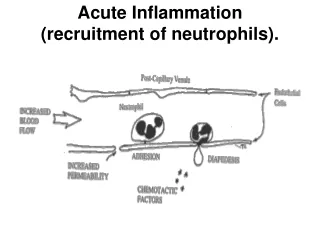
mechanism • Stasis :The loss of fluid and increased vessel diameter lead to slower blood flow, concentration of red cells in small vessels, and increased viscosity of the blood
mechanism • As a result of stasis , blood leukocytes, principally neutrophils, accumulate along the vascular endothelium ( margination). • Rolling and adhesion: Leukocytes then adhere to the endothelium through adhesion molecules. Mediators: adhesion molecules Rolling ( unstable adhesion) through selectins firm adhesion through integrins.
mechanism • Then migrate through the vascular wall into the interstitial tissue (exravasation) or called transmigration or diapedesis Mediators :Chemokines act on the adherent leukocytes and stimulate the cells to migrate through interendothelial spaces toward the chemical concentration gradient, that is, toward the site of injury or infection where the chemokines are being produced The leukocyte moves by extending filopodia that pull the back of the cell in the direction of extension.
mechanism • leukocytes pierce the basement membrane, probably by secreting collagenases, and enter the extravascular tissue • chemotaxis, which is defined as locomotion oriented along a chemical gradient. After exiting the circulation, leukocytes emigrate in tissues toward the site of injury. Both exogenous and endogenous substances can act as chemoattractants. Exogenous agents are bacterial products, Endogenous: cytokines( IL8), arachidonic acid metabolites as leukotriene B4, complement system as C5.
during the first 6 to 24 hours : neutrophils predominate • in 24 to 48 hours : neutrophils are replaced by monocytes.
Neutrophils))A :early MI with polymorphonuclrearleucocytesB:old MI with mononuclear leucocytes(lymphocytes ¯ophages
Recognition of Microbes and Dead Tissues by leucocytes through • -Receptors for microbial products • - Receptors for opsonins: Leukocytes express receptors for proteins that coat microbes. The process of coating a particle, such as a microbe, to target it for ingestion (phagocytosis) is called opsonization, and substances that do this are opsonins. Ex. Of opsonins include antibodies, complement proteins, and lectins • - Receptors for cytokines as interferon-γ (IFN-γ)
phagocytosis . • Phagocytosis involves three sequential steps • (1) recognition and attachment of the particle to be ingested by the leukocyte; • (2) engulfment, with subsequent formation of a phagocytic vacuole: After a particle is bound to phagocyte receptors, extensions of the cytoplasm (pseudopods) flow around it, and the plasma membrane pinches off to form a vesicle (phagosome) that encloses the particle • The phagosome then fuses with a lysosomal granule, resulting in discharge of the granule's contents into the phagolysosome
(3) killing or degradation of the ingested material: Microbial killing is accomplished largely by: • reactive oxygen species (ROS) and reactive nitrogen species. • Neutrophil granules enzymes, such as elastase
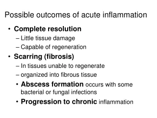
Outcomes of Acute Inflammation • 1-Completeresolution:Resolution involves removal of cellular debris and microbes by macrophages, and resorption of edema fluid by lymphatics • 2-Healing by connective tissue replacement (fibrosis). Causes are: -substantial tissue destruction, - when the inflammatory injury involves tissues that are incapable of regeneration, or -when there is abundant fibrin exudation in tissue or serous cavities (pleura, peritoneum) that cannot be adequately cleared. In all these situations, connective tissue grows into the area of damage or exudate, converting it into a mass of fibrous tissue—a process also called organization. • 3-Progression of the response to chronicinflammation
Morphologic Patterns of Acute Inflammation • SEROUS INFLAMMATION • Serous inflammation is marked by the outpouring of a thin fluid that may be derived from the plasma or from the secretions of mesothelial cells lining the peritoneal, pleural, and pericardial cavities. Accumulation of fluid in these cavities is called an effusion
FIBRINOUS INFLAMMATION • A fibrinousexudate develops when the vascular leaks are large or there is a local procoagulant stimulus (e.g., cancer cells). fibrin is formed and deposited in the extracellular space • A fibrinousexudate is characteristic of inflammation in the lining of body cavities, such as the meninges, pericardium and pleura. • Histologically, fibrin appears as an eosinophilic meshwork of threads or sometimes as an amorphous coagulum. • Fibrinous exudates may be removed by fibrinolysis and clearing of other debris by macrophages.
SUPPURATIVE OR PURULENT INFLAMMATION; ABSCESS • production of large amounts of pus or purulent exudate consisting of neutrophils, liquefactive necrosis, and edema fluid • Ex. acute appendicitis • Bacteria produce suppuration called pyogenic (pus forming) • Abscesses are localized collections of purulent inflammatory tissue caused by suppuration buried in a tissue, an organ, or a confined space