Download
1 / 30
300 likes | 321 Vues
Development of a silicon detector for photon counting to be used in dual energy radiography in the range 18-40KeV. Digital Mammography X ray tests Phantoms spectra Bilateral Project (ICM-tube X-ray). Digital Mammography. Vantages: Better resolution Image manipulation Image Inversion

E N D
Development of a silicon detector for photon counting to be used in dual energy radiography in the range 18-40KeV • Digital Mammography • X ray tests • Phantoms spectra • Bilateral Project (ICM-tube X-ray)
Digital Mammography Vantages: • Better resolution • Image manipulation • Image Inversion • Identification of other tissues • Reduction of time in studies (half)
Characteristics of SD • Position identification • Energy 8-30 KeV • Spatial resolution 50-100 um • Fast electronics • Low noise • Low dose
Key system issues Fully parallel signal processing for all channels. Only binary (yes/no) information is extracted from each strip. Data from each channel must be stored in a local buffer for the whole measurement period. Silicon strip detector 64 PC computer bond wires
0.6 1.0 cm2 First prototype (1xRX64) detector RX64
Detector Tests • Calibration through an internal pulse to obtain gain and noise estimation of the system • Calibration through radiactive sources to obtain gain, noise and the absolute energy scale • Detector test using a X ray tube at CINVESTAV and a mammograph at Hospital Juárez • Obtain spectra with phantoms made in laboratory and an official phantom (ACR).
Hospital Juárez-México Fis. Enrique Gaona (UAM-Xochimilco)
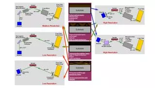
Scanning system – 2D image • Advantages compare to • 2D-systems: • reduced scatter background, • small number of the electronics • channel. scanning slit
Activities in Mexico 2004 • Caracterization and tests in hospital by A. Leyva (posdoc ALFA). • Simulation of the system by a portuguese student (ALFA). • Test with ACR phantom by Roxana de la Mora (Bil. Proj. Cuba)
Phantom 1 (First tests) Colimator: 250um Steps: 2 Time: 3600s/step Cd-109 (22,24keV)
Semiconductor diode Image Colimador: 1mm Pasos: 16 Tiempo: 600s/paso Cd-109 (22,24keV)
Internal image LED Collimator: 1mm Steps: 26 Time: 3600s/step Cd-109 (22,24keV)
Structures Images with Cd-109 Collimator: 0.5mm Steps: 26 Time: 2400s/step Cd-109 (22,24keV)
ACR Phantom The ACR phantom has structures that simulate microcalcifications of several sizes.
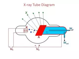
BEDE X ray Tube Anode: Cu, window: Be ICM Project
Mass profile (13) X ray tube mass 1mm
Characteristics of mammographin Hospital Juárez • Elscint, made in Israel • Model MAM-CH225 • X ray tube model : XRHAVA • Range: 22-35 kV • Automatic adjustment of current • Anodo-filter: Mo/Mo
Detail 12 and phantom 1First results mass 2mm Phantom 1 Time:0.8s, 28kV, 90 mAs
Biological Tissue (uterus) Analysis in progress…
Activities-Ricardo • 1(*)- Estudio de: • a) Interacción de las radiaciones con la materia. • b) Detección de las radiaciones ionizantes. • d) Detectores semiconductores de silicio • 2- Calibración 2 chips RX64. • a) Determinación de la linealidad de los contadores. • b) Determinar Ampl DAC y Shaper DAC para el sistema. • c) Medición de la ganancia, offset y el ruido.
Activities… • 3- Calibración energética del sistema de detección empleando fuentes patrones radioactivas. • 4- Obtención de imágenes de objetos en el laboratorio • 5- Simulacion del sistema con Geant
Activities done so far • 1- Study of radiation interaction with matter (silicon) (*) • 2- Study simulation programs (Litrani, Geant) (*) • 3- Measurement of gain, noise and offset through calibration pulse and radiation sources. (see report) • 4- Programs to obtain mean and sigma of data (developed in C) (*) * Should be in the report
Future activities… • Most of the measurement done sofar with edge-on configuration • Poisson test. • Biológical tissues measurementwith both front and edge on configutations. • Measurements with X ray tube and Mammograph.