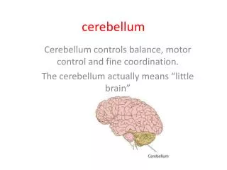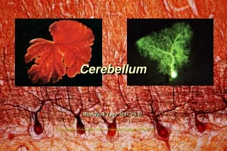
Cerebellum
Cerebellum. Dr. Safaa. Objectives Identify the major lobes and regions of cerebellum. Summarize the structure of the cerebellar cortex Identify the deep cerebellar nuclei and their connections.


Cerebellum
E N D
Presentation Transcript
Cerebellum Dr. Safaa
Objectives • Identify the major lobes and regions of cerebellum. • Summarize the structure of the cerebellar cortex • Identify the deep cerebellar nuclei and their connections. • List the afferent and efferent connections of the cerebellum and their arrangement in cerebellar peduncles. • Describe the major functions of the cerebellum and how each side of the cerebellum controls the ipsilateral side of the body. • Explain the effects of lesions of cerebellum and motor disorder associated with cerebellar lesions.
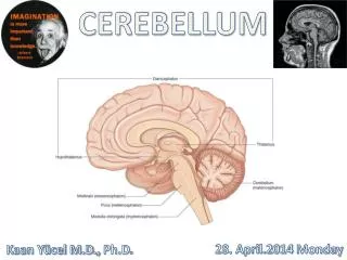
Gross Appearance of the Cerebellum • The cerebellum is situated in posterior cranial fossa • Covered superiorly by tentorium cerebelli. • Lies posterior to the fourth ventricle, the pons, and the medulla oblongata. • It consists of two cerebellar hemispheres joined vermis. • The cerebellum is connected to the posterior aspect of the brainstem by: • Superior cerebellar peduncles (midbrain). • Middle cerebellar peduncles (pons). • Inferior cerebellar peduncles (medulla oblongata).
The cerebellum is divided into three main lobes: • Anterior lobe which is separated from the middle lobe primary fissure. • Middle (posterior) lobe which is situated between the primary and uvulonodular fissures. • Flocculonodular lobe. which is situated posterior to the uvulonodular fissure A deep horizontal fissure separates the superior from the inferior surfaces.
Structure of the Cerebellum • The cerebellum is composed of: • Outer cortex of gray matter. • Inner white matter. • Three masses of gray matter (intracerebellar nuclei) are embedded in the white matter.
The cerebellar cortex is formed of folds called folia. • Each fold or folium contains a core of white matter covered superficially by gray matter. • A section made through the cerebellum parallel with the median plane has a branched appearance, called the arbor vitae. • The gray matter of the cortex may be divided into three layers: • (1) Molecular layer (stellate& basket cells). • (2) Purkinje cell layer (Purkinje cells) . • (3) Granular layer (granular & golgicells).
Intracerebellar Nuclei: 1.Dentate: The largest of the cerebellar nuclei. It has the shape of a crumpled bag 2. Emboliform:Is ovoid 3. Globose: consists of one or more rounded cell groups 4. Fastigial:Lies near the midline in the vermis. • Afferents to these nuclei from: • Inhibitory from Purkinje cells. • Excitatory from climbing and mossy fibers. • Efferents from these nuclei form the cerebellaroutflow in the superior and inferior cerebellar peduncles.
White Matter: • The white matter is made up of three groups of fibers: • (1) Intrinsic. • (2) Afferent. • (3) Efferent.
Afferent fibers (mossy & climbing fibers): • From cerebral cortex(Conveys control from cerebral cortex) : • Corticopontocerebellar through pontine nuclei in middle peduncle & terminates as mossy fibers tocerebellar cortex. • Cerebroreticulocerebellar through reticular formation in inferior and middle peduncles& terminates as mossy fibers to cerebellar cortex. • Cerebro-olivocerebellarthrough inferior olivary nuclei in inferior peduncle & terminates as climbing fibers to cerebellar cortex.
2. From spinal cord (conveys information from muscles and joints): • Anterior spinocerebellarfrom nucleus dorsalis (Clarke's column) majority crossing and enter cerebellum through superior peduncle & terminates as mossy fibers tocerebellar cortex. • Posterior spinocerebellarfrom nucleus dorsalis (Clarke's column) NOT crossing and enter cerebellum through inferior peduncle & terminates as mossy fibers to cerebellar cortex.
3.Cuneocerebellar(Conveys information from muscles and joints of upper limb)originate in nucleus cuneatus of medulla oblongata NOT crossing and enter cerebellum through inferior peduncle& terminates as mossy fibers to cerebellar cortex. 4. Vestibular nerve (conveys information of head position and movement) originate in inner ear NOT crossing and enter cerebellum through inferior peduncle& terminates as mossy fibers to flocculonodular lobe. 5. In addition, cerebellum receives small bundles of afferent fibers from red nucleus & tectum.
Efferent fibers (to red nucleus, thalamus, vestibular complex, and reticular formation) : • Purkinje cell axons synapse with all cerebellar nuclei(fastigial, globose, emboliform, and dentate). • From dentate, emboliform, and globose nuclei through the superior cerebellar peduncle. • From fastigial nucleus leave through the inferior cerebellar peduncle.
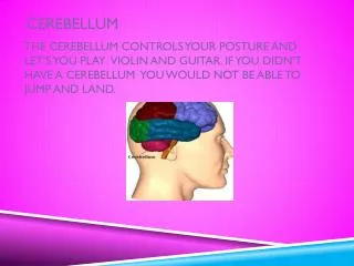
Each cerebellar hemisphere influences the voluntary muscle tone on the same side of the body(ipsilateral). • The cerebellum has no direct neuronal connections with the lower motor neurons but exerts its influence indirectly through the cerebral cortex and brainstem.