Robust Optic Disk Segmentation from Colour Retinal Images
230 likes | 490 Vues
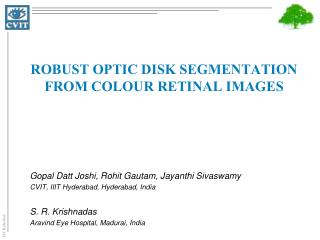
Gopal Datt Joshi, Rohit Gautam, Jayanthi Sivaswamy CVIT, IIIT Hyderabad, Hyderabad, India S. R. Krishnadas Aravind Eye Hospital, Madurai, India. Robust Optic Disk Segmentation from Colour Retinal Images. Optic Disk (OD) Segmentation. Changes in OD are indicative for Glaucoma

Robust Optic Disk Segmentation from Colour Retinal Images
E N D
Presentation Transcript
Gopal Datt Joshi, Rohit Gautam, Jayanthi Sivaswamy CVIT, IIIT Hyderabad, Hyderabad, India S. R. Krishnadas Aravind Eye Hospital, Madurai, India Robust Optic Disk Segmentation from Colour Retinal Images
Optic Disk (OD) Segmentation • Changes in OD are indicative for Glaucoma • Direct ophthalmoscope is used to assess changes • a subjective OD parameterisation • Retinal image analysis is a valuable aid for the objective assessment • OD segmentation is a fundamental task
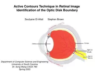
Challenges • irregular disk shape • blood vessel occlusions • ill defined boundaries • atrophy presence • regions around OD with similar image characteristics • inter and intra image variations Smooth region transition Vessels Atrophy region Optic disk boundary
OD SegmentationState of the Art Template Matching Gradient based Active Contour Region based Active Contour • Based on • Shape • Intensity • Lalonde et al. 2001 • Chrastek et al. 2002 • Abdel-Ghafar et al. 2007 • Pallawala et al. 2004 • Snakes • Mendels et al. 1999 • Lowell et al. 2004 • Li et al. 2003, 2004 • Wong et al. 2008 • Novo et al. 2009 • Juan et al. 2007 • Chan Vese (C-V) Active contour • Yandong et al. (2006): • Joshi et al. (2010) • Sensitive to local gradients • To handle: • Impose shape prior • (Fixed or Learned) • Evolution on selective curve points • Sensitive to contour initialisation Fixed shape assumption… Advantages & Limitations >> next
Region-based Active Contour • Advantages: • lower sensitivity to contour initialisation and noise • feasibility of segmentation of color images even in the absence of gradient-defined boundaries • better ability to capture concavities of objects • Attempts with Chan Vese (C-V) model • Yandong et al. (2006): with a circularityconstraint • Joshi et al. (2010): with no shape constraint
C-V model: Limitations Example of smooth region transition Erroneous segmentations where the object cannot be easily distinguished in terms of global statistics expert C-V model Example of high atrophy
Objective • To achieve consistent and robust segmentation Strategy • by integrating local statistics • to improve sensitivity to the slowly varying gradient boundaries • by integrating information from multiple image feature channels • to differentiate the OD region from atrophy regions
Basic Chan Vese (C-V) Model • Consider a vector valued image where in the image domain • The C-V model defines the energy functional as: contour ‘C’ outside inside c+ and c- are two constants to approximate the image intensity inside and outside of the contour C. λ and µ are weights for the fitting and the regularizing terms, respectively
Proposed Model • Underlying assumption in C-V model • image consists of statistically homogeneous regions • lacks in handling inhomogeneous objects • The basic idea is • local instead of global statistics • to extend the scope of the model • information from multiple feature channel • to bring robustness against distraction near OD region
Localisation of C-V model outside region The redefined energy for a point ‘x’ where, h+ and h- are two constants that approximate region intensities inside and outside a contour, near the point x inside region defines a local image domain around a point x with in the radius of r x and y denotes two points in the image I. This energy is minimum when a point is exactly on the boundary
Multi-feature Channels Integration outside Integration of multiple feature channels to the model as: inside
Original Colour Image Red colour space • gives a better discriminating representation of image regions • make model robust to the distractions found near the OD boundaries Texture Space- 2 Texture Space- 1
Energy minimisation over all Points • The integral of over all points ‘x’ is minimised to obtain entire object boundary, defined as: • An equivalent level-set formulation is defined for curve evolution* * Refer to the article for the details of level-set formulation.
Experimentations • Dataset: 138 images of size (2896 x 1944 pixels) • collected from Aravind Eye Hospital, Madurai • Ground Truth: from 3 eye experts • we derive an average boundary to compensate inter-observer variability • Comparison: • With two known active contour models • Gradient vector flow (GVF) • Basic C-V model • Contour initialisation and pre-processing are kept same • To only assess the strength of individual model
Results Input Initialisation GVF C-V model Proposed model Expert Marking Method’s Result
Inter Observer Variability • This is due to • level of clinical experience • comfort level with the marking tool The proposed method has better consensus with the average marking White: Proposed method; Other: Experts
Evaluation Metrics • Region-based: pixel wise segmentation accuracy • Boundary-based: boundary localisation accuracy • Let Cg be the boundary marked by the expert and Co be the boundary obtained by the method
Results on 8 Difficult Images Average signed boundary Distance Under segmentation Over-segmentation
Results on 138 images F-score Average Boundary Distance
Conclusion • We presented a novel, active contour model to achieve robust OD segmentation • Contributions: • the scope of C-V model is extended • by localising energy functional • robust active contour model • by integrating multiple image feature channels • suitable for various OD shapes • no shape prior used
Thanks GopalDatt Joshi PhD Student CVIT, IIIT Hyderabad email: gopal@reserach.iiit.ac.in
ROI selection and Contour initialisation • The red colour plane of retinal image was chosen • it gives good definition of OD region • The vessel points are identified and masked using standard vessel segmentation technique • We perform localisation and initialisation steps together by performing circular Hough transform on the gradient map • Identified circle radius and center point are used to get initialisation for the contour