Download
1 / 28
280 likes | 846 Vues
LOCALIZATION. Cerebellum NMJ Brachial Plexus, Median, Radial, and Ulnar nerves. Cerebellum [17 cm x 120cm]. Anatomy Relation 3 Lobes Functional 4 deep nuclei fastigial Interposed x 2 dentate Topography Blood supply. Vermis Ms mvt of the axial body

E N D
LOCALIZATION Cerebellum NMJ Brachial Plexus, Median, Radial, and Ulnar nerves
Cerebellum [17 cm x 120cm] • Anatomy • Relation • 3 Lobes • Functional • 4 deep nuclei • fastigial • Interposed x 2 • dentate • Topography • Blood supply
Vermis Ms mvt of the axial body The intermediate zone distal portion of the limbs & face The lateral zone [has no topographic representation] Input motor & premotor areas of the frontal cortex & somatosensory & sensory ass. area of the parietal cortex overall planning & coordination of rapid sequential mvts Incoordination
Functions • Timing, in rapid progression • Intensity of muscle contraction • Continuously updated info. • Makes corrective adjustments when actual mvt unfavorably compares with the intended • Performs most of the damping function • Aids the C.Cx in planning next mvt smooth progression • Ballistic mvts [typing, guitar] loss of automaticity • Control the interplay b/n agonist & antagonists, +synergists & fixators • Extramotor predictive function:- rate of progression of auditory and visual phenomenon
Afferent pathways Brain • Corticopontocerebellar Pathway to lat zone • Olivocerebellar tract to all areas • Vestibuloc. fibers floculonod lobe & Fastigial N. • Reticulocerebellr fibers vermis Periphery • Dorsal spinocerebellar tract vermis & IM zone same side • From ms spindles and somatic • Ventral spinocerebellar tract [fastest] • SCP Both side • By Motor signals arriving at AHC [“efference copy of the ant horn motor drive]
Major Efferent pathways • Midlinefastigial N. medullary & pontine fastigiobulbar & C.reticular tracts • Equilibrium apparatus & vestibular N. Equil. • Reticular formation postural attitude • IM zoneinterposed NC. reticular & C. .olivery fs • Thalamus Cx, BG, to the red N and RF • Coordinate reciprocal contraction of limbs ms • Lateral zonedentate N. thalamus Cx Dentatothalamic & Dentatorubral fs • coordination of sequential motor activity
Summary: It controls motor fn at 3 level • The Vestibulocerebellum [archicerebellum] • floculonodular + adjacent vermis • Equilibrium and postural mvts • The Spinocerebellum [paleocerebellum] • IM zone comparison interposed N corrective signal • Smooth coordinated mvt of agonist and antagonist ms of distal limb for acute purposeful patterned mvt • Damping & ballistic mvt fn • Cerebrocerebellum [Neocerebellum] • Lateral zone & dentate N • Extreme incoordin. of cx purposeful mvts of hands,fingers, & feet & of speech apparatus • Planning & Timing of sequential mvts • Extramotor predictive function
Clinical Abnormalities • Small lesion / !/2 removed • Imbalance and Ataxia • Truncal ataxia wide based gait • Not lateralized +/- Symmetric nystagmus toxic, metabolic, inflammatory or neurodegenerative dis. • Asymmetric ataxia structural dis. Ischemia, tumor or mass lesion • Visual cues • C. limb ataxia • Dysmetria • Dysdiadochokinesia • Tremor • Past pointing / rebound • C. nystagmus • Hypotonia • Dysarthria
Cerebellar Syndromes • The Rostral Vermis Sundrome [ant.lobe] • Wide-based stance and gait • Ataxia of Gait vs. heel-to shin • Arm coordination Nl/impaired • Hypotonia/nystagmus/dysarthria infrequent Cerebellar cortical degeneration of Chr. Alcoholics • Ant & sup vermis
Caudal Vermis Synd [Floculonodular & post lobe] • Axial dysequilibirium & staggering gait • Little or no limb ataxia • +/- nystagmus &rotated head posture • S/S of Inc ICP medulloblastoma
Cerebellar Hemispheric Synd • Mvts requiring fine coord. [ms controlled by the precentral cx] Infarcts, neoplasm, and abscesses • Pancerebellar Syndrome • Trunk, limbs, cranial ms • Infectious and parainfectious, Hypoglycemia, hyperthermia, paraneoplastic d/o, and other toxic-metabolic d/os
Syndromes of cerebellar infarction • Thrombotic or embolic • Limb & gait ataxia, dysarthria, nyst, & altered mental sttus • With/without BS and 4th vent compression • Large occiputal headache vetigo, N/V, gait unsteadiness and dysarthria, obst hydrocephalus • neck stiffness, • Herniation CR sx VS. MB compression • PICA, SCA or both • Border zone not easily localizable • 47 pts: cardiac arrest [4%], Atheroma or hypercoagulable state [20%], Large art VB occlusive dis. [34%], Brain embolism [23%], unknown mechanism [19%]
Approach • Hx Duration, Neurologic Sx, Alcohol, Nutritional… • Exam –sensory/ motor • Evaluation for Vit. B12 def. • Imaging studies
Neuromuscular Junction Dis. • Fatigable/ ptosis, diplopia or bulbar weakness • Proximal weakness • Normal mental fn, • Fluctuating • Ms tone/Reflexes/Atrophy • Most often gradual
Major causes of intermittent generalized weakness are: • Electrolyte disturbances • Ms disorders [channelopathies, metabolic defects] • NMJ disorders [MG, LEMS] • Myopathic proximal and are rarely limited to the limbs • Proximal weakness of 2/4 limbs ms, less commonly, NMJ or AHC • Myopathy pelvic or shoulder girdle ms • NM disorders such as MG Proximal weakness ass with ptosis,diplopia or bulbar weakness & fluctuating in severity during the day • The proximal weakness of AHC dis asymmetric • Numbness is absent in all • sensation is intact
Myasthenia Gravis • Autoantibody anti AchR • The most common [1 in 7500]] • Fatigable weakness • The cranial ms Early • Diplopia, ptosis, Chewing, speech, regurgitation, aspiration • 85% generalized • DTR & sensation preserved • Response to Tx, RNS
DDX • Neurasthenia nonorganic • Drug induced myasthenia • LEMS presynaptic/ auto Ab against Ca++ channels[85%] • Often ass with malignancy • Hyper-/hypothyroidism inc. myasthenic weakness • Grave’s disease VS. ocular MG • Botulism pupil and Incremental RNS • Intracranial mass lesion • Congenital myasthenic syndrome
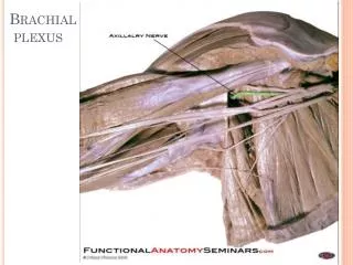
The Brachial Plexus • C5-T1 to the entire UL + shoulder and neck ms • root, trunk, division, cords and branches. • 5 major nerves: Axillary, Radial [posterior], Musculocutaneous [Lateral], Ulnar [medial] and Median [lateral + Medial] • Plexopathy Diffuse focused study of PNs • Radiculopthy sensation is spared • Most B.P regional involvement localizing to the specific region in the B.P 1st step then easy
General rules • Weakness in a “myotomal” pattern C8 to T1 mslower trunk/medial cord C5 and C6 ms upper trunk/lateral cord isolated to a single nr is unlikely to be of plexus origin, Inv’t of ms innervated by Radial & axill nrPost cord Isolated middle trunkunheard C7 radiculopathy Fixed sensory loss extending into the medial forearm lower trunk/medial cord plexopathy; sensory loss extending into the lat forearm upper trunk/lateral cord plexopathy
Weakness of the serratus anterior radiculopathy or a component • Specific disorders of the brachial plexus • Downward movement of the shoulder results in an upper trunk disorder (Erb's palsy) • hyperabduction causes a lower trunk injury (Klumpke's palsy). • Lower trunk plexopathy by compression TOS
Upper trunk plexopathies may ff surgical intervention in the region of the neck. • Lower trunk plexopathies also can occur after chest-splitting cardiothoracic surgery. • Compression by tumors/masses lower trunk plexopathies from “Pancoast tumors” • Backpack palsy —patients often present with arm/shoulder weakness after wearing a backpack for a prolonged period of time, suggestive of a predominantly upper trunk lesion. Some sensory loss is also present.
RADICULOPATHY • Structural spine disease of the cervical spine remains one of the most frequent problems affecting the upper extremities. • In the young individual with an acute herniated disc, severe radiating pain, sensory loss, and weakness in muscles of the myotome of the affected nerve root can be present. • In the elderly- acute herniation of discs is uncommon; cervical spondylosis 2o to disc degeneration, calcification,… more frequent --.they often present present with more diffuse pain, sensory loss, and weakness. involvement of multiple myotomes • Head turning, coughing, or sneezing may exacerbate symptoms, regardless of age. • C7 radiculopathies 70%. C6 n 19 to 25 %; C8 in 4 to 10 %; and C5 in 2 %.
C5 radiculopathy — Sry lossproximal lat arm. Weakness shoulder muscles including deltoid and biceps brachii . Biceps reflex- lost • C6 radiculopathy — Sry loss lat forearm and digit 1 of the hand. Weaknessbiceps and brachioradialis, deltoid, pronator teres, and triceps minimally. Biceps and brachioradialis reflex lost • C7 radiculopathy — Sry loss digits 2 to 4, with digit 3 most affected. Weakness triceps , wrist flexors, pronator teres, and wrist extensors. Triceps reflex lost. • C8 radiculopathy — Sry loss digit 5, medial hand, and medial forearm. Weakness finger flexors, thumb abduction, interossei, and finger extensors.
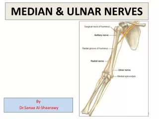
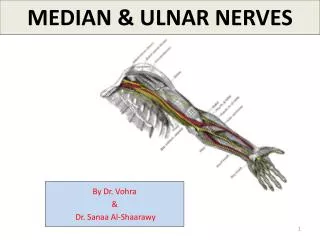
MEDIAN NERVE SYNDROMES • Pronator teres syndrome [rare] • Entrapment in the proximal forearm physically active . • Forearm pain and sry loss over entire lateral palm. Sry loss over the thenar eminence typical, Vs. from carpal tunnel syndrome • Anterior interosseous neuropathy br around the elbow. innervates the flexor pollicis longus, the deep flexors of digits 2 and 3, and pronator quadratus. not sry; O/E: cannot make a standard "O"
ULNAR NERVE SYNDROMES • At the elbow — Ulnar neuropathy is the 2nd most common compression neuropathy. sry loss and paresthesias over digits 4 and 5. & worsened grip and clumsiness. plus weakness in finger and wrist flexion. A prominent Tinel's sign • Epicondylar groovethe Tardy ulnar palsy • Entrapment as it enters the cubital tunnel • At the wrist same finding – finger flexers
RADIAL NERVE SYNDROMES • At the spiral groove ffs prolonged pressure . "Saturday night palsy" • O/E the triceps is OK the wrist, finger extensors, and brachioradialis are weak. Sry loss over the dorsum of the hand, possibly extending up the posterior forearm. Thumb abduction is affected as abductor pollicis longus is a radial-innervated muscle. • Posterior interosseous neuropathy — nerve branches off just proximal to the elbow and innervates the extensor muscles of the forearm O/E medial deviation of the wrist