Animal Development and Phylogeny
760 likes | 809 Vues
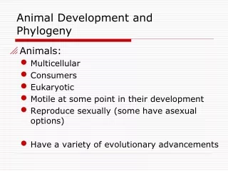
Animal Development and Phylogeny. Animals: Multicellular Consumers Eukaryotic Motile at some point in their development Reproduce sexually (some have asexual options) Have a variety of evolutionary advancements. The Animal Family Tree.

Animal Development and Phylogeny
E N D
Presentation Transcript
Animal Development and Phylogeny • Animals: • Multicellular • Consumers • Eukaryotic • Motile at some point in their development • Reproduce sexually (some have asexual options) • Have a variety of evolutionary advancements
The Animal Family Tree • The most primitive animals are conglomerations of cells with little specialization and no true tissues (sponges) • More advanced animals have cells organized into distinct tissues (Eumetazoa) • Diploblastic organisms have only 2 tissue layers (cnidarians and ctenophorans) • Triploblastic organisms have 3 tissue layers (look in a mirror)
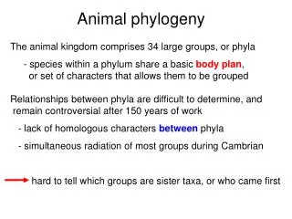
Asymmetry Sponge Figure 32-5 No plane of symmetry Radial symmetry Jellyfish Multiple planes of symmetry Bilateral symmetry Lizard Single plane of symmetry Posterior Anterior
Animal Activities-minor grade and NOTES • Use the animal activities to determine evolutionary patterns in Kingdom Animalia. • Complete each one on your own paper. Use a textbook, online notes, online resources and previous knowledge. • The last question for each sections is a Thinkable this is the question that makes the connection between information and knowledge
A word about Germ layers • “Germ” layers refers to the 3 layers of tissues in most animals. The layers are present at gastrulation during embryonic development • Ectoderm is the outermost layer of cells. It gives rise to the nervous system, skin, hair and nails • Mesoderm is the middle layer of cells and is the most versatile. It becomes the skeleton, muscles, inner layer of skin, visceral lining, fatty tissues, and circulatory system • Endoderm is the innermost layer of cells. It gives rise to the gut and organs associated with digestion and excretion
Introduction to Kingdom Animalia • Evolution • Work through the first two activities: Symmetry and Tissue Layers • 30 minutes.
Why Symmetry? • Most primitive organisms are asymmetric, slightly more advanced are radially symmetric, and the most advanced are bilaterally symmetric • Why? • Segmentation is tied to bilaterally symmetry. • Organisms with bilateral symmetry tend to have more advanced features such as sensory organs. Cephalization!!!!
The Animal Family Tree • Animals with true tissues and bilateral symmetry are considered the most advanced and classified into three groups: • Specialized tissues and basic organs but no body cavity (acoelomates) • Still more advanced organisms develop a body cavity which is unlined (pseudocoelomates) • The most advanced organisms develop a body cavity lined in mesoderm (coelomates) • Body cavities allow organisms to form sections for specialized organs and organ systems. • This leads to segmentation=Evolutionary Money!
Acoelomates have no enclosed body cavity. Skin (from ectoderm) No coelom Muscles, organs (from mesoderm) Figure 32-6 Gut (from endoderm) Pseudocoelomates have an enclosed body cavity partially lined with mesoderm. Skin (from ectoderm) Pseudocoelom Muscles, organs (from mesoderm) Gut (from endoderm) Coelomates have an enclosed body cavity completely lined with mesoderm. Skin (from ectoderm) Coelom Muscles, organs (from mesoderm) Gut (from endoderm)
Family Tree Continued • The coelomates are further divided into two groups: • Protostomes-”proto”=first, “stome”=mouth, • Deuterostomes-”deutero”=second, “stome”=mouth • Groups are based on the fate of the Blastopore during gastrulation • Protostomes are all invertebrates. • Deuterostomes are echinoderms and chordates.
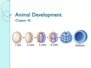
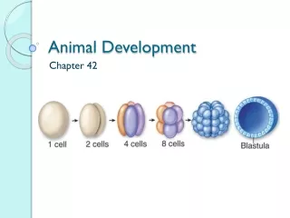
PROTOSTOMES DEUTEROSTOMES 2-cell stage Cleavage (zygote undergoes rapid divisions, eventually forming a mass of cells) Figure 32-8 4-cell stage 8-cell stage Spiral cleavage Radial cleavage Gastrulation (mass of cells formed by cleavage is rearranged to form gut and embryonic tissue layers) Longitudinal section Mouth Pore becomes mouth Pore becomes anus Anus Coelom formation (body cavity lined with mesoderm develops) Gut Gut Coelom Mesoderm Mesoderm Block of solid mesoderm splits to form coelom Mesoderm pockets pinch off of gut to form coelom Cross section
gellates als Amoebozoa Choanofla Fungi Anim Figure 32-10 Animalia Bilateria Deuterostoma Protostoma Ecdysozoa Lophotrochozoa (sea stars, sand dollars) (segmened worms) (insects, spiders, crustaceans) (snails, clams, squid) Choanoflagellates (jellyfish, sea anemeones) Platyhelminthes Echinodermata Acoelomorpha (roundworms) (vertebrates, ascidians) (comb jellies) Ctenophora Arthropoda Nematoda Chordata (flatworms) (sponges) Porifera Annelida (rotifers) Mollusca Cnidaria Rotifera (acoels) Radial symmetry (in adults) Segmentation Pseudocoelom Segmen- tation Acoelom Segmen- tation Pseudocoelom Growth by molting Deuterostome development Protostome development Coelom Triploblasty (origin of mesoderm) Phylogenetic tree based on similarities and differences in the DNA sequences of several genes from various animal phyla. The bars along the branches indicate when certain morphological traits originated Bilateral symmetry and cephalization Radial symmetry Diploblasty (ectoderm and endoderm) Epithelial tissue Multicellularity
Kindgom Animalia Activity Continued…… • Write three columns on your paper: Know Think I Know Don’t Know • Sort the following terms into an appropriate column for YOU: Bilateria, Radiata, Acoelomates, Coelomates, Pseudocoleomates, Protostome, Eumtazoa, Parazoa, Choanoflagellate, Deuterostome
Animal Activity Continued….. • Define the terms in your “Don’t Know” Column using your notes, neighbor, electronic devices or textbook. • Start with #3 on the activity sheet. Use the pieces to help you sort and resort but write the final product in your notes. • 15 minutes
Animal Classification/Review • Molecular data continues to change our views on how animals are grouped into phyla. The bilaterally symmetric animals are particularly messy to classify • There are some points of agreement with respect to classification: • All animals share a common ancestor • Sponges are the base of the animals family tree • Eumetazoa is a clade of animals with true tissues (cnidaria and ctenophora, formerly coelenterata) • Most animal phyla belong to the Bilateria clade and are organized based on the presence of a coelom. • Chordates and some other phyla belong to the clade Deuterostoma
Major Invertebrate Phyla • Sponges were formerly called “Porifera” and are organisms that have the following characteristics: • Suspension feeding (capturing food from the water as it travels through the body • Pores on the outer surface pull in water and send it out through the spongocoel and it’s main opening, the osculum • All are hermaphroditic • Have a few specialized cells but no tissues: • Choanocytes-collar cells that are flagellated for feeding • Amoebocytes-mobile cells that have pseudopods and carry nutrients around the body • These are now split into 2 phyla: • Calcarea • Silicea
Figure 32-26 Pseudoceratina crassa
Eumetazoans • This is a clade, consisting of 2 major phyla of diploblastic organisms: • Cnidaria (Includes: jellyfish, hydra, sea anemones, etc) • Radially symmetrical • Tissue layers (2 distinct-epidermis, gastrodermis)-mesoglea in between (jelly) • 2 forms-medusa (mouth down, free-swimming), and polyp (mouth up, sessile) • Stinging nematocysts for defense and predation (inside the cnidocytes) • 1st organisms with a nervous system (primitive-nerve net, no central control) • Food enters the mouth and broken down. Nutrients from the food are absorbed by the surrounding cells and wastes are expelled from the mouth (2-way digestive tract) • Ctenophora (Comb Jellies) • Look like jellyfish, but move with cilia on their bodies • No cnidocytes/nematocysts, instead use colloblast secretions to catch and hold onto prey • Actually have a nervous control structure called the Apical Organ at one end of the body (sounds like a brain to me)
Figure 32-27 Medusae float near the water surface. Polyps attach to substrates. Aurelia aurita Aurelia aurita
Motile larval anemone Sessile adult anemone Figure 32-18
Figure 32-3 Cnidarians and ctenophores are diploblastic. Cnidaria include hydra, jellyfish, corals, and sea pens (shown). Ctenophora are the comb jellies. Ectoderm Endoderm This dark blue comb jelly… …has just swallowed this white comb jelly
Figure 32-4 Mouth Captured prey will be transferred to mouth Tentacles Tubular body Basal disk
Figure 32-28 Pleurobrachia pileus Rows of cilia Sticky tentacles
Acoelomates • Also called the flatworms b/c they have no body cavity and a flattened body • First organisms with bilateral symmetry and cephalization • Organisms with a two-way digestive tract or none at all • No need for lungs or gills because of the flat body plan (O2 exchange via diffusion) • Water-living or parasitic
Figure 33-13 Turbellarians are free living. Cestodes are endoparasitic. Trematodes are endoparasitic. Taenia species Pseudoceros ferrugineus Dicrocoelium dendriticum
Rotifers • Small, freshwater organisms with a ciliated crown • Have an alimentary canal with 1-way digestion • Some species can reproduce via parthenogenesis and are all female, while others have males only for the purpose of reproduction
Rotaria rotatoria Figure 33-12 Corona
Mollusca • Bilaterally symmetric • Muscular Foot (ventral) • Rasping organ called the Radula • Coelomates • Open circulatory system • Primitive kidneys • Gills or primitive lungs • Several ganglia with a more complex nervous system • Examples include snails, slugs, chitons, limpets, bivalves (clams, oysters, mussels, scallops), chambered nautilis, squid, octopus
Mollusc body plan (internal view) Figure 33-7b Gill Muscular “foot” Visceral mass (internal organs and external gill) Mantle (secretes shell)
Scallops live on the surface of the substrate and suspension feed. Lima scabra Figure 33-15 Most clams burrow into soft subtrates and suspension feed. Water out Food particles Water in Gill Siphons Gills are thin structures for gas exchange. They also trap food particles as water passes through them. Cilia move the particles to the mouth Foot
Snails have a single shell, which they use for protection. Maxacteon flammea Figure 33-16 Land slugs and sea slugs (nudibranchs) lack shells. Chromodoris geminus Bright colors warn potential predators of presence of toxins
Figure 33-17 Tonicella lineata
Figure 33-18 Octopus dofleini
Nematodes • Have round bodies (pseudocoel) • Both free-living and parasitic • Ex: hook worm, Ascaris, pinworm, trichina worm, dog heartworm • Often have complex life styles w/intermediate hosts • Often have male and female forms with dimorphism
Figure 33-21 Strongyloides species Nematodes
Annelids • 1st organisms with segmentation (metamerism) • Closed circulatory system but gas exchange occurs via osmosis • Double nerve cord, ganglia, lateral nerves in each segment (metamere) • Taste, tactile, light sensation • Bilaterally symmetric • Head (prostomium) and an anus-bearing terminal portion • Hydrostatic skeleton in each segment
Figure 33-14 Most leeched live in freshwater. Most polychaetes are marine. Most oligochaetes are terrestrial. Alvinella pompejana Paranais litoralis Hirudo medicinalis Chaetae
Arthropods • Arthro=jointed, pod=foot, all have jointed appendages • Exoskeleton made of chitin (a protein) and sometimes calcium carbonate • Metamorphosis • Bilateral symmetry, open circulation, nervous system like that of annelids • Have gills, air tubes, or book gills • Defined body segments and developed sensory organs.
Figure 33-7a Arthropod body plan (external view) Tagma Thorax Abdomen Head Jointed limbs Exoskeleton (covers body) Segmented body
Figure 33-23 Spider, showing general chelicerate features Mites are ectoparasitic. Dolomedes fimbriatus Dermatophagoides species Posterior region Anterior region Chelicerae
Deep-sea lobster Red barnacle Figure 33-24 Enoplometopus occidentalis Tetraclita species Barnacles secrete their own shells Carapace Fiddler crab Uca vocans Compound eyes on stalks
Echinoderms • Non-metameric adult with radial symmetry • Larvae are bilaterally symmetric • No head or brain, circular ring and radial nerves • Skeleton of embedded ossicles (calcium carbonate) within the dermis • Pedicellariae for catching and moving food • Water vascular system with tube feet for locomotion • One-way digestive tract (sometimes with eversible stomach) • Dermal branchae also help with vascularization • Usually separate sexes • Ex: sea stars, sea lillies, sea urchins
Figure 34-2 Echinoderm larvae are bilaterally symmetric. Adult echinoderms are radially symmetric.
Figure 34-3 Echinoderms have a water vascular system. Podia project from the underside of the body. Opening to exterior Tube foot Podia Podia
Figure 34-21 Sea urchin Sand dollar Echinus tylodes Dendraster excentricus Teeth at center of underside