Download
1 / 62
630 likes | 765 Vues
Nucleic acids, including DNA and RNA, are crucial carriers of genetic information in cells. DNA contains the instructions that dictate cellular structure and function, guiding growth, division, and protein synthesis. RNA plays a vital role in translating these instructions into functional proteins. Nucleotides, the building blocks of nucleic acids, are linked through phosphodiester bonds, forming long chains. This overview explores the structures of DNA and RNA, their nucleotide compositions, and key processes such as replication, transcription, and translation critical for genetic expression.

E N D
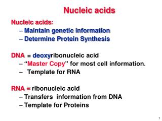
Nucleic Acids Nucleic Acids are the chemical carriers of a cell’s genetic information • Deoxyribonucleic acid (DNA) • Holds the information that determines the nature of a cell • Controls cell growth and division • Directs biosynthesis of the enzymes and other proteins required for cellular functions • Ribonucleic acid (RNA) • Nucleic acid derivatives such as ATP are involved as phosphorylating agents in many biochemical pathways • Several important coenzymes, including NAD+, FAD, and coenzyme A, have nucleic acid components
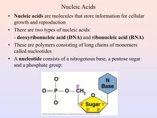
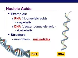
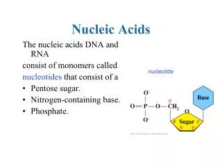
24.1 Nucleotides and Nucleic Acids Nucleic acids are biopolymers • Composed of nucleotides which are joined together to form a long chain • Nucleotide • Composed of nucleosides bound to a phosphate group • Nucleoside • Composed of an aldopentose sugar linked through its anomeric carbon to the nitrogen atom of a heterocyclic purine or pyrimidine base
Nucleotides and Nucleic Acids DNA • Sugar component is 2′-deoxyribose (the prefix 2′-deoxy indicates that oxygen is missing from the 2′ position of ribose) • Contains four different amino bases • Two substituted purines (adenine and guanine) • Two substituted pyrimidines (cytosine and thymine) RNA • Sugar component is ribose • Contains adenine, guanine, and cytosine • Thymine is replaced by a closely related pyrimidine base called uracil
Nucleotides and Nucleic Acids The pyrimidines and purines found in DNA and RNA
Nucleotides and Nucleic Acids Structures of the four deoxyribonucleotides
Nucleotides and Nucleic Acids Structures of the four ribonucleotides
Nucleotides and Nucleic Acids • In naming and numbering nucleotides, positions on the sugars are given a prime superscript to distinguish them from positions on the amine base • DNA and RNA differ dramatically in size • Molecules of DNA have molecular weights up to 75 billion • Molecules of RNA are much smaller, containing as few as 21 nucleotides, and have a molecular weight as low as 7,000
Nucleotides and Nucleic Acids • Nucleotides are linked together in DNA and RNA by phosphodiester bonds between the phosphate group at C5′ on one nucleotide and the 3′-hydroxyl group of the sugar of another nucleotide • C3′ is one free hydroxyl group at the end of the nucleic polymer (the 3′ end) • C5′ is another free hydroxyl group at the other end of the nucleic polymer (the 5′ end) • Sequence of nucleotides in a chain is described by starting at the 5′ end and identifying the bases in order of occurrence (using G, C, A, T or U)
24.2 Base Pairing in DNA: The Watson- Crick Model Samples of DNA isolated from different tissues of the same species have the same proportions of heterocyclic bases Samples of DNA from different species often have greatly different proportions of bases • Composition of human DNA • 30% each of adenine and thymine • 20% each of guanine and cytosine • Composition of the bacterium Clostridium perfringens • 37% each of adenine and thymine • 13% each of guanine and cytosine
In 1953, James Watson and Francis Crick proposed the secondary structure of DNA DNA under physiological conditions consists of two polynucleotide strands Strands run in opposite directions and coil around each other in a double helix The helix is 20 Å wide The two strands are complementary and are held together by hydrogen bonds between specific pairs of bases A with T C with G Base Pairing in DNA: The Watson-Crick Model
There are 10 base pairs per turn Each turn is 34 Å in length The two strands of the double helix coil in such a way that two kinds of “grooves” result A major groove 12 Å wide A minor groove 6 Å wide The grooves are lined with hydrogen bond donors and acceptors A variety of flat, polycyclic aromatic molecules are able to slip sideways, or intercalate, between the stacked bases An organism’s genetic information is stored as a sequence of deoxyribonucleotides strung together in the DNA chain Base Pairing in DNA: The Watson-Crick Model
Base Pairing in DNA: The Watson-Crick Model “Central dogma of molecular genetics” • The function of DNA is to store information and pass it to RNA • The function of RNA is to read, decode, and use the information received from DNA to make proteins • Three fundamental processes take place: • Replication – process by which identical copies of DNA are made so the information can be preserved and handed down to offspring • Transcription – the process by which the genetic messages are read and carried out of the cell nucleus to ribosomes, where protein synthesis occurs • Translation – the process by which the genetic messages are decoded and used to synthesize proteins
Worked Example 24.1Predicting the Complementary Base Sequence in Double-Stranded DNA What sequence of bases on one strand of DNA is complementary to the sequence TATGCAT on another strand?
Worked Example 24.1Predicting the Complementary Base Sequence in Double-Stranded DNA Strategy • Remember that A and G form complementary pairs with T and C • Go through the sequence replacing A by T, G by C, T by A, and C by G • Remember that the 5′ end is on the left and the 3′ end is on the right in the original strand
Worked Example 24.1Predicting the Complementary Base Sequence in Double-Stranded DNA Solution Original: (5′) TATGCAT (3′) Compliment: (3′) ATACGTA (5′) or (5′) ATGCATA (3′)
24.3 Replication of DNA Replication • An enzyme-catalyzed process • Begins with a partial unwinding of the double helix • Bases become exposed • New nucleotides line up on each strand in a complementary manner (A with T and C with G) • Two new strands begin to grow • Each new strand is complementary to its old template strand • Two identical DNA helices are produced • The process is described as semiconservative replication because each of the new DNA molecules contains one old strand and one new strand
Replication of DNA A representation of semiconservative DNA replication
Replication of DNA Addition of nucleotides to the growing chain • Takes place in the 5′→3′ direction • Catalyzed by DNA polymerase • Key step is the addition of a nucleoside 5′-triphosphate to the free 3′-hydroxyl group of the growing chain, with loss of a diphosphate leaving group
Replication of DNA Both new strands are synthesized in the 5′→3′ direction • They cannot be made in exactly the same way • One strand must have its 3′ end near the point of unraveling (the replication fork), while the other strand has its 5′ end near the replication fork • The complement of the original 5′→3′ strand is synthesized continuously in a single piece • The compliment of the original 3′→5′ strand is synthesized discontinuously in small pieces that are often then linked by DNA ligases
24.4 Transcription of DNA RNA • Similar to DNA but contains ribose instead of deoxyribose and uracil instead of thymine • Three primary kinds • Messenger RNA (mRNA) carries genetic messages from DNA to ribosomes, • Small granular particles in the cytoplasm of a cell where protein synthesis takes place • Ribosomal RNA (rRNA) complexed with protein provides the physical makeup of the ribosomes • Transfer RNA (tRNA) transports amino acids to the ribosomes where they are joined together to make proteins
Transcription of DNA Genetic information in DNA is contained in segments called genes • Each gene consists of a specific nucleotide sequence that encodes a specific protein • Conversion of DNA information into proteins begins with transcription of DNA to mRNA
Transcription of DNA Promoter site • A specific base sequence found within a DNA chain typically consisting of around 40 base pairs located upstream (5′) of the transcription start site • Consists of two hexameric consensus sequences, one located 10 base pairs upstream and the second located 35 base pairs upstream from the beginning of the coding region • Signals the beginning of a gene • Other base sequences signal a stop near the end of the gene
Transcription of DNA Transcription • The process by which genetic information encoded in DNA is read and used to synthesize RNA in the nucleus of the cell • Several turns of the DNA double helix unwind, forming a “bubble” and exposing the bases of the two strands • Ribonucleotides line up in the proper order by hydrogen bonding to their complementary bases on DNA • Bond formation occurs in the 5′ 3′ direction • The growing RNA molecule unwinds from DNA
Transcription of DNA Only one of the two DNA strands is transcribed into mRNA • The strand that contains the gene is called the coding strand, or sense strand • The strand that gets transcribed is called the template strand, or antisense strand • The RNA molecule produced during transcription is the complement of the DNA antisense strand and is therefore a copy of the DNA coding strand (except T has been replaced with U)
Transcription of DNA Genes are not continuous segments of the DNA chain • A gene begins in an exon, a small section of DNA • Genes are interrupted by noncoding sections called introns • Genes take up again further down the chain in another exon The final mRNA molecule results after the noncoded sections are cut out and the remaining pieces are spliced together • 90% of human DNA seems to be made up of introns • 10% of DNA contains coding instructions
24.5 Translation of RNA: Protein Biosynthesis Primary cellular function of mRNA • Direct biosynthesis of the thousands of diverse peptides and proteins required by an organism • The mechanics of protein biosynthesis take place on ribosomes, small granular particles in the cytoplasm of a cell that consist of about 60% ribosomal RNA and 40% protein • The specific ribonucleotide sequence in mRNA forms a codon that determines the order in which amino acid residues are joined • Each codon consists of a sequence of three ribonucleotides that is specific for a given amino acid • The series UUC on mRNA is a codon directing incorporation of the amino acid phenylalanine into the growing protein • 64 possible triplets of the four bases in RNA • 61 code for specific amino acids • 3 code for chain termination
Translation of RNA: Protein Biosynthesis Translation • The process by which the genetic information transcribed from DNA onto mRNA is read by tRNA and used to direct protein synthesis • There are 61 different tRNAs, one for each of the 61 codons that specifies an amino acid • A typical tRNA is single-stranded and cloverleaf-shaped • On the middle leaf it contains an anticodon, a sequence of three ribonucleotides complementary to the codon sequence • Contains about 70 to 100 ribonucleotides • Bonded to a specific amino acid by an ester linkage through the 3′ hydroxyl on ribose at the 3′ end of the tRNA
Translation of RNA: Protein Biosynthesis • The codon sequence UUC present on mRNA is read by a phenylalanine-bearing tRNA having the complementary anticodon base sequence GAA • Nucleotide sequences are written in the 5′→3′ direction so the sequence in an anticodon must be reversed
Translation of RNA: Protein Biosynthesis • Successive codons on mRNA are read • Different tRNAs bring the correct amino acids into position for enzyme-mediated transfer to the growing peptide • When synthesis of the proper protein is completed, a “stop” codon signals the end • The protein is released from the ribosome
Worked Example 24.2Predicting the Amino Acid Sequence Transcribed from DNA What amino acid sequence is coded by the following segment of a DNA coding strand? (5′) CTA-ACT-AGC-GGG-TCG-CCG (3′)
Worked Example 24.2Predicting the Amino Acid Sequence Transcribed from DNA Strategy • The mRNA produced during translation is a copy of the DNA coding strand • Each T replaced by U • The mRNA has the sequence (5′) CUA-ACU-AGC-GGG-UCG-CCG (3′)
Worked Example 24.2Predicting the Amino Acid Sequence Transcribed from DNA Solution • Leu-Thr-Ser-Gly-Ser-Pro
24.6 DNA Sequencing Methods for sequencing immense DNA chains • First step in sequencing • Cleave the DNA chain at known points to produce smaller pieces, done through the use of restriction endonucleases • More than 3500 restriction enzymes are known • About 200 restriction enzymes are commercially available • Each different restriction enzyme cleaves a DNA molecule at a point in the chain where a specific base sequence occurs • The restriction enzyme AluI cleaves between G and C in the four-base sequence AG-CT • (5′-AGCT-(3′) sequence is that same as its complement (3′)-TCGA-(5′) when both are read in the same 5′→3′ direction
DNA Sequencing Two methods of DNA sequencing are available • The Maxam-Gilbert method • Uses chemical techniques • Sanger dideoxy method • Uses enzymatic reactions • The more commonly used of the two • Method responsible for sequencing the entire human genome of 2.9 billion base pairs • In commercial sequencing instruments, the dideoxy method begins with a mixture of the following: • The restriction fragment to be sequenced
DNA Sequencing • A small piece of DNA called a primer, whose sequence is complementary to that on the 3′ end of the restriction fragment • The four 2′-deoxyribonucleoside triphosphates (dNTPs) • Very small amounts of the four 2′ ,3′-dideoxyribonucleoside triphosphates (ddNTPs), each of which is labeled with a fluorescent dye of a different color (A 2′ ,3′ -dideoxyribonucleoside triphosphate in one in which both 2′ and 3′ –OH groups are missing from ribose)
DNA Sequencing • DNA polymerase is added to the mixture • A strand of DNA complementary to the restriction fragment begins to grow from the end of the primer • Most of the time only normal deoxyribonucleotides are incorporated into the growing chain • Sometimes a dideoxyribonucleotide is incorporated • When this occurs, DNA synthesis stops because the chain end no longer has a 3′ hydroxyl group for adding further nucleotides • The product • Consists of a mixture of DNA fragments of all possible lengths, each terminated by one of the four dye-labeled dideoxyribonucleotides • Mixture is then separated according to the size of the pieces by gel electrophoresis
DNA Sequencing • The identity of the terminal dideoxyribonucleotide in each piece – and thus the sequence of the restriction fragment – is identified by noting the color with which it fluoresces
24.7 DNA Synthesis Synthesis of short DNA segments, called oligonucleotides or oligos • A nucleotide has multiple reactive sites that must be selectively protected and deprotected at the proper times • Coupling of the four nucleotides must be carried out in the proper sequence • Automated DNA synthesizers allow the fast and reliable synthesis of DNA segments up to 200 nucleotides in length • A protected nucleotide is covalently bonded to a solid support • One nucleotide at a time is added to the growing chain by the use of a coupling reagent • After the final nucleotide has been added, all the protecting groups are removed and the synthetic DNA is cleaved from the solid support
DNA Synthesis Step 1 Attachment of a protected deoxynucleoside to a silica (SiO2) support • Done through an ester linkage to the 3′ –OH group of the deoxynucleoside • Both the 5′ –OH group on the sugar and free –NH2 groups on the heterocyclic bases must be protected • The deoxyribose 5′ –OH is protected as its p-dimethoxytrityl (DMT) ether
DNA Synthesis • Adenine and cytosine bases are protected by benzoyl groups • Guanine is protected by an isobutryl group • Thymine requires no protection
DNA Synthesis Step 2 Removal of the DMT protecting group by treatment with dichloroacetic acid in CH2Cl2 • Reaction occurs by an SN1 mechanism • Reaction proceeds rapidly due to the stability of the tertiary, benzylic dimethoxytrityl cation
DNA Synthesis Step 3 Coupling of the polymer-bonded deoxynucleoside with a protected deoxynucleoside containing a phosphoramiditegroup, R2NP(OR)2, at the 3′ position • Takes place in the polar aprotic solvent acetonitrile • Requires catalysis by the heterocyclic amine tetrazole • Yields a phosphite, P(OR)3
DNA Synthesis Step 4 Oxidation • Phosphite product is oxidized to a phosphate by treatment with iodine in aqueous tetrahydrofuran in the presence of 2,6-dimethylpyridine • The cycle is repeated until oligonucleotide chain of the desired sequence is built • Deprotection • Coupling • Oxidation
DNA Synthesis Step 5 Final step • Removal of all protecting groups • Cleavage of the ester bond holding the DNA to the silica • All reactions are done at the same time by treatment with aqueous NH3 • Purification by electrophoresis yields the synthetic DNA
24.8 The Polymerase Chain Reaction Polymerase chain reaction (PCR) • A method for amplifying small amounts of DNA to produce larger amounts • Invented by Kary Mullis in 1986 • PCR produces multiple copies of a given DNA sequence • Makes it possible to obtain several micrograms (1 ug = 10-6 g; about 1011 nucleotides) in a few hours when starting from less than 1 picogram of DNA with a chain length of 10,000 nucleotides (1 pg = 10-12 g; about 100,000 molecules)
The Polymerase Chain Reaction Taq polymerase • The key to the polymerase chain reaction • A heat-stable enzyme isolated from the thermophilic bacterium Thermus aquaticus found in a hot spring in Yellowstone National Park • Able to take a single strand of DNA that has a short, primer segment of complementary chain at one end and then finish constructing the entire complementary strand • Overall process takes three steps
The Polymerase Chain Reaction The polymerase chain reaction
The Polymerase Chain Reaction Step 1 Denaturation of the double-stranded DNA • The double-stranded DNA is heated in the presence of: • Taq polymerase • Mg2+ ion • The four deoxynucleotide triphosphate monomers (dNTPs) • A large excess of two short oligonucleotide primers of about 20 bases each • Each primer is complementary to the sequence at the end of one of the target DNA segments • Double-stranded DNA denatures at a temperature of 95 ºC, spontaneously breaking apart into two single strands