Bradycardia
Bradycardia. National Pediatric Nighttime Curriculum Written by Jennifer Everhart, MD Lucile Packard Children’s Hospital. Learning Objectives. Recognize common pediatric cardiac bradyarrhythmias Appreciate the clinical significance of bradycardia

Bradycardia
E N D
Presentation Transcript
Bradycardia National Pediatric Nighttime Curriculum Written by Jennifer Everhart, MD Lucile Packard Children’s Hospital
Learning Objectives • Recognize common pediatric cardiac bradyarrhythmias • Appreciate the clinical significance of bradycardia • Initiate management of bradyarrhythmias in the inpatient setting
Case #1 • 9 year old boy admitted for asthma exacerbation, noted to have heart rate of 55. • What do you think is going on? • What is the first thing you would assess in your evaluation of this patient? • What work-up would you do? • How would you treat this child? Thaler 2003
Bradyarrhythmias - Symptoms • Symptoms: • General: altered LOC, fatigue, lightheadedness, dizziness, syncope • Hemodynamic instability: hypotension, poor end-organ perfusion, respiratory distress/failure, sudden collapse
Bradyarrhythmias - Causes • General causes: • 1º: Abnormal pacemaker/conduction system (congenital or postsurgical injury), cardiomyopathy, myocarditis • 2º: Reversible Hs & Ts: • – Hypoxia – Hypotension – Heart block – H+ ions (acidosis) – Hypothermia – Hyperkalemia – Trauma (head) – Toxins/drugs (cholinesterase inhibitors, Ca++ channel blockers, β-adrenergic blockers, digoxin, central α2 adrenergic agonists, opioids)
Case #2 A 7 year old boy with a history of neonatal intraventricular hemorrhage underwent a ventriculoperitoneal shunt revision this afternoon. You are notified by the nursing staff that he is having heart rates down to the 50’s. Teaching file – Courtesy of Wilson King, MD
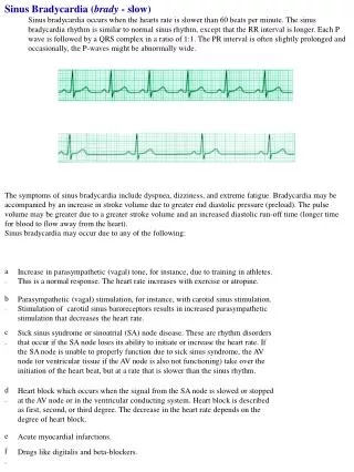
Sinus Bradycardia • Can be a physiologic consequence of decreased metabolic demand (ie: while sleeping) or increased stroke volume (ie: athletes) • Other potential causes include: • Endocrine: hypothyroidism, hypoglycemia • Neurologic: seizures or head trauma causing increased vagal tone; ↑ICP • Ingestion: Beta-blockers, Ca++ channel blockers, digoxin, antiarrhythmics • Hypothermia • Infectious: Sepsis • Sinus bradycardia is almost never primarily cardiac in origin in pediatrics.
Sinus Node Block/Arrest • Caused by absent pacemaker activity in the sinus node with subsidiary pacemakers in the atrium, AV junction/node, or ventricles initiating depolarization: • Atrial escape: Late P wave, different P wave morphology • Junctional escape: Narrow-complex, +/- retrograde P waves • Idioventricular escape: Wide-complex, typical rate 30-40 beats/min Thaler 2003
Case #3 • A 4 month old boy was transferred from the CVICU earlier in the day, following an uncomplicated repair of his VSD. His nurse notifies you that his rhythm on the cardiac monitor looks odd. • What do you think is going on? • What is the first thing you would assess in your evaluation of this patient? • What work-up would you do? • How would you treat this child? Yanowitz, 2006
AV Blocks 1st degree heart block 2nd degree heart block, Mobitz I 2nd degree heart block, Mobitz II 3rd degree heart block Ralston et al, 2006
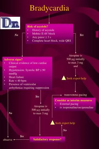
Bradyarrhythmias – Management • Stable patients: • 12 lead EKG • Consult cardiology • Unstable patients: • ABCs • PALS Pediatric Bradycardia Algorithm • Address reversible causes (Hs & Ts) • Consider labs • Ie: blood gas, chemistry panel, digoxin level if applicable Kleinman et al 2010
Take home points • When evaluating a patient with a bradyarrhythmia, the first step is always to address clinical/hemodynamic stability: airway, breathing, circulation. Further management is guided by the PALS Pediatric Bradycardia Algorithm. • Sinus bradycardia is rarely due to primary cardiac pathology in children – reversible causes should be sought and addressed. • Atrial, junctional, and idioventricular escape rhythms are the result of subsidiary pacemakers initiating depolarization in the event of sinus node failure. • 1st, 2nd, and 3rd degree AV blocks vary in etiology and clinical significance. 1st degree and 2nd degree Mobitz Type I are often minimally symptomatic and even self-resolving. 2nd degree Mobitz Type II is more symptomatic, can progress to 3rd degree, and may require pacemaker. 3rd degree is the most symptomatic, and usually requires pacemaker.
References • Key References for independent study: • Kleinman ME, et al. Part 14: Pediatric Advanced Life Support: 2010 American Heart Association Guidelines for Cardiopulmonary Resuscitation and Emergency Cardiovascular Care. Circulation 2010;122;S876-S908. • Ralston M, et al. PALS Provider Manual. American Heart Association, 2006. • Additional References used to prepare this presentation: • 12 Lead EKG Interpretation Part #2, nursingpub.com. • American Heart Association. 2005 American Heart Association (AHA) Guidelines for Cardiopulmonary Resuscitation (CPR) and Emergency Cardiovascular Care (ECC) of Pediatric and Neonatal Patients: Pediatric Advanced Life Support. Pediatrics 2006;117;e1005-1028. • Emergency Medicine Education Online, www.emedu.org. • Fleisher GR, et al. Textbook of Pediatric Emergency Medicine 5th Edition. Lippincott Williams & Williams, 2006. • Thaler MS. The Only EKG Book You’ll Ever Need4th Edition. Lippincott Williams & Williams, 2003. • Yanowitz FG. The Alan E. Lindsay ECG Learning Center in Cyberspace. University of Utah School of Medicine, 2006. • Zaoutis LB and Chiang VW. Comprehensive Pediatric Hospital Medicine. Mosby Elsevier, 2007.
Questions You are cross-covering a previously healthy 15 year old boy admitted for new-onset polyarthritis. His nurse calls to notify you that his heart rate has been in the high-40s and low-50s overnight. • List 5 potential causes of his bradycardia:
Question 2 As you are walking through the oncology unit, you are approached by a nurse who asks you to quickly evaluate her patient with bradycardia. Upon entering the room, you find the 4 year old girl in bed, nonresponsive, poorly perfused but with intact pulses, with a heart rate of 35 on her cardiac monitor. Think through how you would approach this patient, and list 8 (or more) interventions you would consider. Try to list them in order of priority/time course – what would your first step be? Are there any interventions that can wait until the patient is stabilized?
Question 3 3. Which of the following statements regarding bradyarrhythmias in children is correct? • A: Sinus bradycardia is a physiologic consequence of decreased metabolic demand and, if the patient is well-perfused, does not require further investigation • B: Atrial escape, junctional escape, and idioventricular escape rhythms are caused by aberrant conduction through the AV node • C: MobitzI and Mobitz II AV blocks have similar clinical significance • D: 3rddegree AV block is the most symptomatic form of heart block, and often requires placement of a pacemaker
Question 4 Identify the following arrhythmias
Question 5 As you are obtaining a history from the mother of a 5 year old girl with known Mobitz Type II 2nd degree heart block who is being admitted for syncopal episodes, the monitor alarms and you notice the heart rate is 32. You glance at the patient and see that she is no longer watching her movie. She does not arouse when her mom calls her name. Her extremities are cool, and her pulses are palpable but faint. Her breathing is unlabored, and her oxygen saturation is 89% on room air. You press the code blue button as the bedside nurse walks in. You and the nurse give oxygen via non-rebreather and begin chest compressions. What is the most appropriate next step? • A: Epinephrine 0.01 mg/kg IV • B: Atropine 0.02 mg/kg IV • C: Check chemistry panel • D: Obtain 12 lead EKG • E: Defibrillate
Question 6 Which of the following is true to AV block? • A: Drugs such as calcium channel blockers, beta blockers, and digoxin can cause 1st and 2nd degree heart block • B: Electrolyte abnormalities such as hyperkalemia can cause acquired 3rd degree heart block • C: 3rddegree heart block is usually asymptomatic, and of little clinical significance if followed closely • D: 1stdegree heart block is a common cause of syncope in adolescents • E: 2nddegree heart block (Mobitz type I) frequently progresses to 3rd degree heart block, and therefore usually necessitates a pacemaker