Download
1 / 30
300 likes | 522 Vues
Chapter 20 The Cardiovascular System: The Heart. Heart pumps over 1 million gallons per year Over 60,000 miles of blood vessels. Heart Location. Anterior surface of heart. Heart is located in the mediastinum area from the sternum to the vertebral column and between the lungs.

E N D
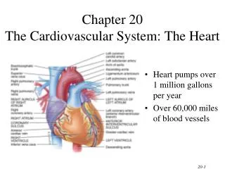
Chapter 20The Cardiovascular System: The Heart • Heart pumps over 1 million gallons per year • Over 60,000 miles of blood vessels
Heart Location Anterior surface of heart • Heart is located in the mediastinum • area from the sternum to the vertebral column and between the lungs
Heart Orientation • Apex - directed anteriorly, inferiorly and to the left • Base - directed posteriorly, superiorly and to the right • Anterior surface - deep to the sternum and ribs • Inferior surface - rests on the diaphragm • Right border - faces right lung • Left border (pulmonary border) - faces left lung
Heart Orientation • Heart has 2 surfaces: anterior and inferior, and 2 borders: right and left
Pericardium • Fibrous pericardium • dense irregular CT • protects and anchors the heart, prevents overstretching • Serous pericardium • thin delicate membrane • contains • parietal layer-outer layer • pericardial cavity with pericardial fluid • visceral layer (epicardium)
Layers of Heart Wall • Epicardium • visceral layer of serous pericardium • Myocardium • cardiac muscle layer is the bulk of the heart • Endocardium • chamber lining & valves
Right Atrium • Receives blood from 3 sources • superior vena cava, inferior vena cava and coronary sinus • Interatrial septum partitions the atria • Fossa ovalis is a remnant of the fetal foramen ovale • Tricuspid valve • Blood flows through into right ventricle • has three cusps composed of dense CT covered by endocardium
Right Ventricle • Forms most of anterior surface of heart • Papillary muscles are cone shaped trabeculae carneae (raised bundles of cardiac muscle) • Chordae tendineae: cords between valve cusps and papillary muscles • Interventricular septum: partitions ventricles • Pulmonary semilunar valve: blood flows into pulmonary trunk
Left Atrium • Forms most of the base of the heart • Receives blood from lungs - 4 pulmonary veins (2 right + 2 left) • Bicuspid valve: blood passes through into left ventricle • has two cusps • to remember names of this valve, try the pneumonic LAMB • Left Atrioventricular, Mitral, or Bicuspid valve
Left Ventricle • Forms the apex of heart • Chordae tendineae anchor bicuspid valve to papillary muscles (also has trabeculae carneae like right ventricle) • Aortic semilunar valve: • blood passes through valve into the ascending aorta • just above valve are the openings to the coronary arteries
Atrioventricular Valves Open • A-V valves open and allow blood to flow from atria into ventricles when ventricular pressure is lower than atrial pressure • occurs when ventricles are relaxed, chordae tendineae are slack and papillary muscles are relaxed
Atrioventricular Valves Close • A-V valves close preventing backflow of blood into atria • occurs when ventricles contract, pushing valve cusps closed, chordae tendinae are pulled taut and papillary muscles contract to pull cords and prevent cusps from everting
Semilunar Valves • SL valves open with ventricular contraction • allow blood to flow into pulmonary trunk and aorta • SL valves close with ventricular relaxation • prevents blood from returning to ventricles, blood fills valve cusps, tightly closing the SL valves
Valve Function Review Which side is anterior surface? What are the ventricles doing?
Valve Function Review Ventricles contract, blood pumped into aorta and pulmonary trunk through SL valves Atria contract, blood fills ventricles through A-V valves
Blood Circulation • Two closed circuits, the systemic and pulmonic • Systemic circulation • left side of heart pumps blood through body • left ventricle pumps oxygenated blood into aorta • aorta branches into many arteries that travel to organs • arteries branch into many arterioles in tissue • arterioles branch into thin-walled capillaries for exchange of gases and nutrients • deoxygenated blood begins its return in venules • venules merge into veins and return to right atrium
Blood Circulation (cont.) • Pulmonary circulation • right side of heart pumps deoxygenated blood to lungs • right ventricle pumps blood to pulmonary trunk • pulmonary trunk branches into pulmonary arteries • pulmonary arteries carry blood to lungs for exchange of gases • oxygenated blood returns to heart in pulmonary veins
Blood Circulation • Blood flow • blue = deoxygenated • red = oxygenated
Coronary Circulation • Coronary circulation is blood supply to the heart • Heart as a very active muscle needs lots of O2 • When the heart relaxes high pressure of blood in aorta pushes blood into coronary vessels • Many anastomoses • connections between arteries supplying blood to the same region, provide alternate routes if one artery becomes occluded
Coronary Arteries • Branches off aorta above aortic semilunar valve • Left coronary artery • circumflex branch • in coronary sulcus, supplies left atrium and left ventricle • anterior interventricular art. • supplies both ventricles • Right coronary artery • marginal branch • in coronary sulcus, supplies right ventricle • posterior interventricular art. • supplies both ventricles
Coronary Veins • Collects wastes from cardiac muscle • Drains into a large sinus on posterior surface of heart called the coronary sinus • Coronary sinus empties into right atrium
Conduction System of Heart Coordinates contraction of heart muscle.
Autorhythmic Cells Cells fire spontaneously, act as pacemaker and form conduction system for the heart SA node cluster of cells in wall of Rt. Atria begins heart activity that spreads to both atria excitation spreads to AV node AV node in atrial septum, transmits signal to bundle of His AV bundle of His the connection between atria and ventricles divides into bundle branches & purkinje fibers, large diameter fibers that conduct signals quickly Conduction System of Heart
One Cardiac Cycle • At 75 beats/min, one cycle requires 0.8 sec. • systole (contraction) and diastole (relaxation) of both atria, plus the systole and diastole of both ventricles • End diastolic volume (EDV) • volume in ventricle at end of diastole, about 130ml • End systolic volume (ESV) • volume in ventricle at end of systole, about 60ml • Stroke volume (SV) • the volume ejected per beat from each ventricle, about 70ml SV = EDV - ESV
Cardiac Output • Amount of blood pushed into aorta or pulmonary trunk by ventricle • Determined by stroke volume and heart rate • CO = SV x HR • at 70ml stroke volume & 75 beat/min----5 and 1/4 liters/min • entire blood supply passes through circulatory system every minute • Cardiac reserve is maximum output/output at rest • average is 4-5 while athlete is 7-8
Influences on Stroke Volume • Preload (affect of stretching) • Frank-Starling Law of Heart • more muscle is stretched, greater force of contraction • more blood more force of contraction results • Contractility • autonomic nerves, hormones, Ca+2 or K+ levels • Afterload • amount of pressure created by the blood in the way • high blood pressure creates high afterload
Congestive Heart Failure • Causes of CHF • coronary artery disease, hypertension, MI, valve disorders, congenital defects • Left side heart failure • less effective pump so more blood remains in ventricle • heart is overstretched & even more blood remains • blood backs up into lungs as pulmonary edema • suffocation & lack of oxygen to the tissues • Right side failure • fluid builds up in tissues as peripheral edema
Regulation of Heart Rate • Nervous control from the cardiovascular center in the medulla • Sympathetic impulses increase heart rate and force of contraction • parasympathetic impulses decrease heart rate. • Baroreceptors (pressure receptors) detect change in BP and send info to the cardiovascular center • located in the arch of the aorta and carotid arteries • Heart rate is also affected by hormones • epinephrine, norepinephrine, thyroid hormones • ions (Na+, K+, Ca2+) • age, gender, physical fitness, and temperature
Clinical Problems • MI = myocardial infarction • death of area of heart muscle from lack of O2 • replaced with scar tissue • results depend on size & location of damage • Blood clot • use clot dissolving drugs streptokinase or t-PA & heparin • balloon angioplasty • Angina pectoris----heart pain from ischemia of cardiac muscle