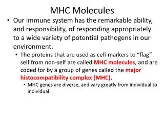
MHC Class I
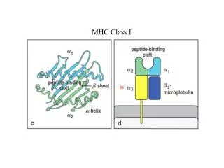
MHC Class I. *. Ends are closed, and 2 -3 pockets at each end. Important Points. Closed ends of class I cleft limit peptide size Anchor residues of peptide fit into class I pockets Peptides bulge or kink Peptide binding motifs predict T cell epitopes

MHC Class I
E N D
Presentation Transcript
Important Points • Closed ends of class I cleft limit peptide size • Anchor residues of peptide fit into class I pockets • Peptides bulge or kink • Peptide binding motifs predict T cell epitopes • MHC polymorphisms located in peptide binding cleft Changes what peptides can bind class I • Change in anchor residue of peptide changes its ability to bind class I
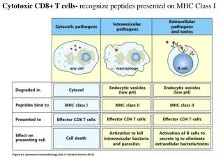
MHC Class I Expression • Wide-spread - Almost all nucleated cells • Exceptions include sperm • Not on human RBC - Used by Malaria • Cytokines increase class I gene transcription including interferons, tumor necrosis factor-a and lymphotoxin • Numerous pathogens produce molecules that decrease class I expression by various mechanisms
Ends open Fewer pockets Anchor residue of peptide
Important Points • Open ends of class II cleft allow longer peptides to bind • Flat confirmation of peptide • Fewer pockets cause more permissive binding • More difficult to predict T cell epitopes • Polymorphism of class II has same effects as that of class I
b NH2 end COOH end a Most diversity in CDR3 regions - Central location
Superantigens • Protein antigens activate high number of T cell clones • Activated T cell clones express one V • NOT mitogens • Virulence factors of pathogens • Prevent a protective adaptive immune response
Bind outside of cleft Little contact with CDR3
Consequences of MHC Polymorphism • Polymorphic residues located in cleft • Change number & size of pockets • Change shape of walls & floor of cleft • Changes what peptides can bind • Changes T cell repertoire • Increases probability some individuals will be resistant to a pathogen • Decreases possibility pathogen will wipe out a species
GENERAL CONCEPTS • Ability of MHC molecules to bind a given peptide is determined by the shape of the peptide binding cleft and the pockets • Each MHC molecule binds one peptide at a time • But, one MHC molecule can bind 100’s - 1000’s of different peptides • Each molecule is not occupied by the same peptide • Antigen-presenting cells express all MHC class I & class II molecules • Hence, one presenting cell can potentially stimulate 1000’s of different T cell clones
MHC CLASS I MHC CLASS II Polygenic Polygenic Heterodimer Heterodimer A Polymorphic & Constant Chains Two Polymorphic Chains No Equivalent Chains Pair in Trans No Equivalent Preferential Chain Association Ubiquitous Expression Limited Expression Regulated by Cytokines Regulated by Cytokines CD8+ T Cell Restriction CD4+ T Cell Restriction Binds CD8 Binds CD4 Peptide Binding Groove Closed Peptide Binding Groove Open Limits Size of Peptides Longer Peptides Bind Anchor Residues Fit into Pockets Anchor Residues Fit into Pockets Multiple Pockets Fewer Pockets Accurate Prediction of Motifs Prediction More Difficult