Download
1 / 20
210 likes | 253 Vues
Learn about the structure and functions of antibodies, the basic unit being a heterodimer with distinct light and heavy chain components. Discover how antibodies bind to antigens and the significance of monoclonal and polyclonal responses in immunization. Explore the chemical and enzymatic methods that reveal the antibody's critical structure. Overcoming obstacles in antibody sequencing is discussed, along with how immunoglobulins have both variable and constant regions. Start your journey of understanding immunoglobulins here!

E N D
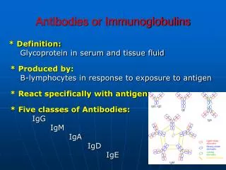
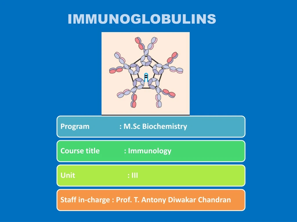
Antibodies are antigen binding proteins present on the B-cell membrane and secreted by plasma cells. Membrane-bound antibody confers antigenic specificity on B cells; antigen-specific proliferation of B-cell clones is elicited by the interaction of membrane antibody with antigen. Secreted antibodies circulate in the blood, where they serve as the effectors of humoral immunity by searching out and neutralizing antigens or marking them for elimination. All antibodies share structural features, bind to antigen, and participate in a limited number of effector functions. The antibodies produced in response to a particular antigen are heterogeneous. Most antigens are complex and contain many different antigenic determinants, and the immune system usually responds by producing antibodies to several epitopes on the antigen.
This response requires the recruitment of several clones of B cells. Their outputs are monoclonal antibodies, each of which specifically binds a single antigenic determinant. Together, these monoclonal antibodies make up the polyclonal and heterogeneous serum antibody response to an immunizing antigen. Basic Structure of Antibodies The first evidence that antibodies were contained in particular serum protein fractions came from a classic experiment by A. Tiselius and E. A. Kabat, in 1939. They immunized rabbits with the protein ovalbumin (the albumin of egg whites) and then divided the immunized rabbits’ serum into two aliquots.
Electrophoresis of one serum aliquot revealed four peaks corresponding to albumin and the alpha (α), beta (β), and gamma (γ) globulins. The other serum aliquot was reacted with ovalbumin, and the precipitate that formed was removed; the remaining serum proteins, which did not react with the antigen, were then electrophoresed. A comparison of the electrophoretic profiles of these two serum aliquots revealed that there was a significant drop in the gamma-globulin peak in the aliquot that had been reacted with antigen. Thus, the gamma-globulin fraction was identified as containing serum antibodies, which were called immunoglobulins, to distinguish them from any other proteins that might be contained in the gamma-globulin fraction.
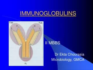
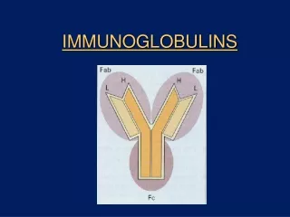
Antibodies Are Heterodimers Antibody molecules have a common structure of four peptide chains. This structure consists of two identical light (L) chains, polypeptides of about 25,000 molecular weight, and two identical heavy (H) chains, largerpolypeptides of molecular weight 50,000 or more. Like the antibody molecules they constitute, H and L chains are also called immunoglobulins. Each light chain is bound to a heavy chain by a disulfide bond, and by such noncovalent interactions as salt linkages, hydrogen bonds, and hydrophobic bonds, to form a heterodimer (H-L). Similar noncovalent interactions and disulfide bridges link the two identical heavy and light (H-L) chain combinations to each other to form the basic four-chain (H-L)2 antibody structure, a dimer of dimers.
Chemical and Enzymatic Methods Revealed Basic Antibody Structure When the γ–globulin fraction of serum is separated into high- and low-molecular weight fractions, antibodies of around 150,000-MW, designated as immunoglobulin G (IgG) are found in the low molecular- weight fraction. In a key experiment, brief digestion of IgG with the enzyme papain produced three fragments, two of which were identical fragments and a third that was quite different . The two identical fragments had antigen-binding activity and were called Fab fragments (“fragment, antigen binding”). The other fragment (MW of 50,000) had no antigen binding activity at all. Because it was found to crystallize during cold storage, it was called the Fc fragment (“fragment, crystallizable”).
Digestion with pepsin, a different proteolytic enzyme, also demonstrated that the antigen-binding properties of an antibody can be separated from the rest of the molecule. Pepsin digestion generated a single 100,000- MW fragment composed of two Fab-like fragments designated the F(ab)2 fragment, which binds antigen. The Fc fragment was not recovered from pepsin digestion because it had been digested into multiple fragments. Mercaptoethanol reduction and alkylation, a chemical treatment that irreversibly cleaves disulfide bonds. If the sample is chromatographed on a column that separates molecules by size following cleavage of disulfide bonds, it is clear that the intact 150,000-MW IgG molecule is, in fact, composed of subunits. Each IgG molecule contains two 50,000-MW polypeptide chains, designated as heavy (H) chains, and two 25,000-MW chains, designated as light (L) chains
Obstacles to Antibody Sequencing Pure Immunoglobulin Obtained from Multiple Myeloma Patients Made Sequencing Possible Light-Chain Sequencing Revealed That Immunoglobulins Have Constant and Variable Regions There were two light chain types, kappa (Ω) and lambda (λ). In humans, 60% of the light chains are kappa and 40% are lambda, whereas in mice, 95% of the light chains are kappa and only 5% are lambda. A single antibody molecule contains only one light chain type, either or , never both. The amino acid sequences of light chains show minor differences that are used to classify light chains into subtypes. In mice, there are three lambda subtypes (1, 2, and 3); in humans, there are four subtypes. Amino acid substitutions at only a few positions are responsible for the subtype differences.
Heavy-Chain Sequencing Revealed Five Basic Varieties of Heavy Chains The length of the constant regions is approximately 330 amino acids for δ, γ, and α, and 440 amino acids for μ and ε. The heavy chains of a given antibody molecule determine the class of that antibody: IgM, IgG,IgA, IgD, or IgE. Each class can have either kappa or lambda light chains. A single antibody molecule has two identical heavy chains and two identical light chains,H2L2, or a multiple (H2L2)n of this basic four-chain structure
Immunoglobulin Fine Structure Immunoglobulins Possess Multiple Domains Based on the Immunoglobulin Fold
HINGE REGION The IgG,IgA and IgD heavy chains contain an extended peptide sequence between the CH1 and CH2 domains that has no homology with the other domains . This region, called the hinge region, is rich in proline residues and is flexible, giving IgG, IgD, and IgA segmental flexibility.As a result, the two Fab arms can assume various angles to each other when antigen is bound. This flexibility of the hinge region can be visualized in electron micrographs of antigen-antibody complexes. For example, when a molecule containing two dinitrophenol (DNP) groups reacts with anti-DNP antibody and the complex is captured on a grid, negatively stained, and observed by electron microscopy, large complexes (e.g., dimers, trimers, tetramers) are seen.
Diversity in the Variable-Region Domains Concentrated in CDRs
Extraction Addition of appropriate amounts of salts, such as ammonium or sodium sulfate, causes precipitation of IgG (1) from all mammals, and can be used for serum, plasma, ascites fluid, and hybridoma culture supernatant. Although such IgG is usually contaminated with other proteins, the ease of these precipitation procedures coupled with the high yield of IgG has led to their wide use in producing enriched IgG preparations. They are suitable for many immunochemical procedures, e.g., production of immunoaffinity columns, and as a starting point for further purification.
About 40% of IgG is precipitated when heparin is added to human plasma at pH 5.4, while only a little IgM and no IgA are precipitated. The precipitation of purified IgG by heparin is pH-dependent and follows a sigmoid curve between pH 7.0 and 5.0. The precipitate has a constant molar ratio heparin: IgG, independent of pH or the amount of heparin that is added. The precipitate does not redissolve at high heparin concentrations. The heavy chains of IgG precipitate also at pH 5.4, but this precipitate redissolves in excess heparin. Light chains do not precipitate and the Fab and Fc fragments are only partly precipitated.
References 1.Immunology by Janis Kuby 2. Cell and molecular Immunology by Abbas and Litchman Thank you