Download
1 / 15
160 likes | 273 Vues
Explore the gross and microscopic anatomy of skeletal muscle, learn about muscle fibers and myofibrils, and discover how muscles work with bones. Understand different muscle types and functions, the ultrastructure of myofibrils, and the role of actin and myosin in muscle contraction.

E N D
Learning Objectives • Learn the gross and microscopic structure of skeletal muscle. • Learn the ultrastructure of a myofibril. • Learn how actin and myosin are arranged within a myofibril.
Muscle and Bone Works Together • Your skeleton is made of bones and joints. • Movement of these components however, is caused by the action of muscles. • Muscles act by pulling bones. • They do so due to receiving nerve impulses. • It is important to realise that muscles are not just slabs of meat, but are highly ordered bundles of fibres. • Glucose, glycogen and fatty acids are sources of energy for muscle contraction.
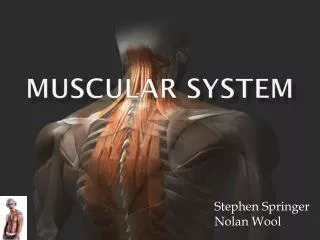
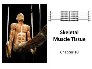
Different Types of Muscle Cardiac muscle is present in the heart. It acts involuntarily. We call it myogenic, because it is able to regulate its own rhythm. • There are actually three types of muscle that can respond to nervous stimulation. Skeletal muscle is the muscle attached to bones. It makes up the vast majority of muscle and can be controlled voluntarily. Smooth muscle is another type of involuntary muscle. This type of muscle is found predominantly in the gut and in the iris of the eye. This lesson focuses on skeletal muscle.
Skeletal Muscle • As mentioned earlier, this muscle type is stimulated voluntarilywhen you want to make movement. • When viewed through a microscope, skeletal muscle has the appearance of being striped. • Skeletal muscle is made up of bundles of muscle fibres. • The picture on the right is highly detailed, but shows the arrangement of muscle fibres into bundles, and the bundles being grouped into a whole muscle.
Skeletal Muscle Whole Muscle • So now you now that muscle is composed of small units that are bundled into progressively larger units. • I.E. • It is the myofibrils that cause skeletal muscle to appear striped (we’ll come on to this later). • It is also the action of the myofibrils that brings about contraction of muscles (next lesson). Bundles of Fibres Muscle Fibres Myofibrils It is useful to think of skeletal muscle as a thick piece of rope. The tiny threads (myofibrils) are very weak individually, but when they are arranged into string (musle fibres), they are stronger. The string can then be wound into thin rope (bundles of fibres), which in turn can be wound into thick rope (whole muscle). The overall structure is immensely strong and fit for the purpose.
Skeletal Muscle Why so nucleated? Remember that each muscle fibre is essentially a massively long cell. It needs nuclei ‘dotted’ along it so that transcription doesn’t just occur in one place. A cell that is so packed with protein, requires protein synthesis to happen all along it. Which is why muscle fibres are multinucleate. • What we haven’t talked about yet are individual ‘muscle cells’. • If muscle consisted of individual cells arranged end-to-end, muscles would be very weak. • Instead, individual cells have been fused together to form the muscle fibres you’ve learnt about. • In a way, the myofibrils are ‘organelles’ within these muscle fibres. • So, because these muscle fibres are such massive cells, they are actually multinucleate – they have many nuclei. nucleus myofibrils Muscle Fibre
Myofibrils There are actually other proteins involved in muscles: Tropomyosinforms a fibrous strand around the two actin filaments. Troponin is a protein involved in muscle contraction (next lesson) • Now that you know muscle fibres are composed of many myofibrils, you need to know these structure of these too. • Myofibrils actually consist of two types of protein filaments: • ACTIN Actin is the thinner of the two filaments that make up a myofibril. It’s actually made up of two strands coiled around each other. • MYOSIN Myosin is a strange-looking filament, which is thicker and consists of rod-shaped fibres that have ‘bulbed’ headsthat project outwards.
Myofibrils • Myofibrils appear striped because of the way that actin and myosin filaments are arranged within in them. • In areas where there is overlapping of actin and myosin, the bands are darker. • In areas where there is no overlapping, the bands appear light. • So basically, actin and myosin filaments are arranged as shown above – these units are known as sarcomeres.
A cross-section of a myofibril, showing actin and myosin filaments, would look like this... There are actually two types of muscle fibres too. SLOW-TWITCH FIBRES These are found in muscles that have to carry out endurance exercise. They therefore have to be adapted to carry out aerobic respiration. FAST-TWITCH FIBRES These contract rapidly and powerfully, but for short periods. They are therefore useful for adapted for intense exercise. These are also adapted for anaerobic respiration.
Sarcomeres Myosin filaments • To understand later how muscles contract, we need to know what happens in the sarcomere, but first we need to understand their structure. • When muscles contract, it involves actin and myosin sliding over each other. • The theory of muscle contraction is therefore known as THE SLIDING FILAMENT MECHANISM. • It therefore follows, that when a muscle contracts, the sarcomere shown above, will slide inwards. Actin filaments
This area appears light, because only thin, actin filaments are present. IT IS CALLED THE I-BAND This area appears dark because there is overlapping of both filaments. IT IS CALLED THE A-BAND. This area, where there are only myosin filaments, is called the H-ZONE Remember that the size of the I and A bands can change, because the filaments slide over each other. This is called the Z-LINE