Download
1 / 43
430 likes | 614 Vues
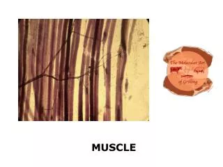
Muscle Overview. The three types of muscle tissue are skeletal, cardiac, and smooth These types differ in structure, location, function, and means of activation. Muscle Similarities. Skeletal and smooth muscle cells are elongated and are called muscle fibers

E N D
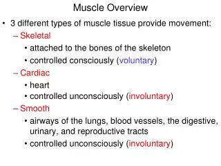
Muscle Overview • The three types of muscle tissue are skeletal, cardiac, and smooth • These types differ in structure, location, function, and means of activation
Muscle Similarities • Skeletal and smooth muscle cells are elongated and are called muscle fibers • Muscle contraction depends on two kinds of myofilaments – actin and myosin • Muscle terminology is similar • Sarcolemma – muscle plasma membrane • Sarcoplasm – cytoplasm of a muscle cell • Prefixes – myo, mys, and sarco all refer to muscle
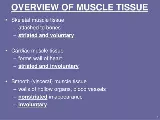
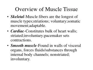
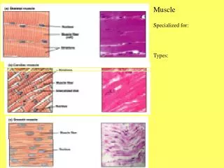
Skeletal Muscle Tissue • Packaged in skeletal muscles that attach to and cover the bony skeleton • Has obvious stripes called striations • Is controlled voluntarily (i.e., by conscious control) • Contracts rapidly but tires easily • Is responsible for overall body motility • Is extremely adaptable and can exert forces ranging from a fraction of an ounce to over 70 pounds
Cardiac Muscle Tissue • Occurs only in the heart • Is striated like skeletal muscle but is not voluntary • Contracts at a fairly steady rate set by the heart’s pacemaker • Neural controls allow the heart to respond to changes in bodily needs
Smooth Muscle Tissue • Found in the walls of hollow visceral organs, such as the stomach, urinary bladder, and respiratory passages • Forces food and other substances through internal body channels • It is not striated and is involuntary
Functional Characteristics of Muscle Tissue • Excitability, or irritability – the ability to receive and respond to stimuli • Contractility – the ability to shorten forcibly • Extensibility – the ability to be stretched or extended • Elasticity – the ability to recoil and resume the original resting length
Muscle Function • Skeletal muscles are responsible for all locomotion • Cardiac muscle is responsible for coursing the blood through the body • Smooth muscle helps maintain blood pressure, and squeezes or propels substances (i.e., food, feces) through organs • Muscles also maintain posture, stabilize joints, and generate heat
Skeletal Muscle • Each muscle is a discrete organ composed of muscle tissue, blood vessels, nerve fibers, and connective tissue
Skeletal Muscle • The three connective tissue sheaths are: • Endomysium – fine sheath of connective tissue composed of reticular fibers surrounding each muscle fiber • Perimysium – fibrous connective tissue that surrounds groups of muscle fibers called fascicles • Epimysium – an overcoat of dense regular connective tissue that surrounds the entire muscle
Skeletal Muscle Figure 9.2a
Skeletal Muscle: Nerve and Blood Supply • Each muscle is served by one nerve, an artery, and one or more veins • Each skeletal muscle fiber is supplied with a nerve ending that controls contraction • Contracting fibers require continuous delivery of oxygen and nutrients via arteries • Wastes must be removed via veins
Skeletal Muscle: Attachments • Most skeletal muscles span joints and are attached to bone in at least two places • When muscles contract the movable bone, the muscle’s insertion moves toward the immovable bone, the muscle’s origin
Skeletal Muscle: Attachments • Muscles attach: • Directly – epimysium of the muscle is fused to the periosteum of a bone • Indirectly – connective tissue wrappings extend beyond the muscle as a tendon or aponeurosis
Microscopic Anatomy of a Skeletal Muscle Fiber • Each fiber is a long, cylindrical cell with multiple nuclei just beneath the sarcolemma • Fibers are 10 to 100 m in diameter, and up to hundreds of centimeters long • Each cell is a syncytium (multinucleate cytoplasm that is not separated into different cells) produced by fusion of embryonic cells
Microscopic Anatomy of a Skeletal Muscle Fiber • Sarcoplasm has numerous glycosomes and a unique oxygen-binding protein called myoglobin • Fibers contain the usual organelles, myofibrils, sarcoplasmic reticulum, and T tubules
Myofibrils • Myofibrils are densely packed, rodlike contractile elements • They make up most of the muscle volume • The arrangement of myofibrils within a fiber is such that a perfectly aligned repeating series of dark A bands and light I bands is evident
Myofibrils Figure 9.3b
Sarcomeres • The smallest contractile unit of a muscle • The region of a myofibril between two successive Z discs • Composed of myofilaments made up of contractile proteins • Myofilaments are of two types – thick and thin
Sarcomeres Figure 9.3c
Myofilaments: Banding Pattern • Thick filaments – extend the entire length of an A band • Thin filaments – extend across the I band and partway into the A band • Z-disc – coin-shaped sheet of proteins (connectins) that anchors the thin filaments and connects myofibrils to one another
Myofilaments: Banding Pattern • Thin filaments do not overlap thick filaments in the lighter H zone • M lines appear darker due to the presence of the protein desmin
Ultrastructure of Myofilaments: Thick Filaments • Thick filaments are composed of the protein myosin • Each myosin molecule has a rod-like tail and two globular heads • Tails – two interwoven, heavy polypeptide chains • Heads – two smaller, light polypeptide chains called cross bridges
Ultrastructure of Myofilaments: Thick Filaments Figure 9.4a,b
Ultrastructure of Myofilaments: Thin Filaments • Thin filaments are chiefly composed of the protein actin • Each actin molecule is a helical polymer of globular subunits called G actin • The subunits contain the active sites to which myosin heads attach during contraction • Tropomyosin and troponin are regulatory subunits bound to actin
Ultrastructure of Myofilaments: Thin Filaments Figure 9.4c
Arrangement of the Filaments in a Sarcomere • Longitudinal section within one sarcomere Figure 9.4d
Sarcoplasmic Reticulum (SR) • SR is an elaborate, smooth endoplasmic reticulum that mostly runs longitudinally and surrounds each myofibril • Paired terminal cisternae (fluid filled sac) form perpendicular cross channels • Functions in the regulation of intracellular calcium levels
Sarcoplasmic Reticulum (SR) • Elongated tubes called T tubules penetrate into the cell’s interior at each A band–I band junction • T tubules associate with the paired terminal cisternae to form triads
Sarcoplasmic Reticulum (SR) Figure 9.5
T Tubules • T tubules are continuous with the sarcolemma • They conduct impulses to the deepest regions of the muscle • These impulses signal for the release of Ca2+ from adjacent terminal cisternae
Triad Relationships • T tubules and SR provide tightly linked signals for muscle contraction • A double zipper of integral membrane proteins protrudes into the intermembrane space • T tubule proteins act as voltage sensors • SR proteins are receptors that regulate Ca2+ release from the SR cisternae
Sliding Filament Model of Contraction • Thin filaments slide past the thick ones so that the actin and myosin filaments overlap to a greater degree • In the relaxed state, thin and thick filaments overlap only slightly • Upon stimulation, myosin heads bind to actin and sliding begins
Sliding Filament Model of Contraction • Each myosin head binds and detaches several times during contraction, acting like a ratchet to generate tension and propel the thin filaments to the center of the sarcomere • As this event occurs throughout the sarcomeres, the muscle shortens
Skeletal Muscle Contraction • In order to contract, a skeletal muscle must: • Be stimulated by a nerve ending • Propagate an electrical current, or action potential, along its sarcolemma • Have a rise in intracellular Ca2+ levels, the final trigger for contraction • Linking the electrical signal to the contraction is excitation-contraction coupling
Nerve Stimulus of Skeletal Muscle • Skeletal muscles are stimulated by motor neurons of the somatic nervous system • Axons of these neurons travel in nerves to muscle cells • Axons of motor neurons branch profusely as they enter muscles • Each axonal branch forms a neuromuscular junction with a single muscle fiber
Neuromuscular Junction • The neuromuscular junction is formed from: • Axonal endings, which have small membranous sacs (synaptic vesicles) that contain the neurotransmitter acetylcholine(ACh) • The motor end plate of a muscle, which is a specific part of the sarcolemma that contains ACh receptors and helps form the neuromuscular junction
Neuromuscular Junction • Though exceedingly close, axonal ends and muscle fibers are always separated by a space called the synaptic cleft
Neuromuscular Junction Figure 9.7 (a-c)
Neuromuscular Junction • When a nerve impulse reaches the end of an axon at the neuromuscular junction: • Voltage-regulated calcium channels open and allow Ca2+ to enter the axon • Ca2+ inside the axon terminal causes axonal vesicles to fuse with the axonal membrane
Neuromuscular Junction • This fusion releases ACh into the synaptic cleft via exocytosis • ACh diffuses across the synaptic cleft to ACh receptors on the sarcolemma • Binding of ACh to its receptors initiates an action potential in the muscle
Destruction of Acetylcholine • ACh bound to ACh receptors is quickly destroyed by the enzyme acetylcholinesterase • This destruction prevents continued muscle fiber contraction in the absence of additional stimuli
Action Potential • A transient depolarization event that includes polarity reversal of a sarcolemma (or nerve cell membrane) and the propagation of an action potential along the membrane
Role of Acetylcholine (Ach) • ACh binds its receptors at the motor end plate • Binding opens chemically gated channels • Na+ and K+ diffuse out and the interior of the sarcolemma becomes less negative • This event is called depolarization