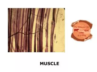
Muscle
Muscle. Chapter 17. Movement. Locomotion Movement of animal from one location to another Repositioning Movement of animal appendages Internal movement Movement of gases, fluids and ingested solids through the animal. Muscle Tissue. Specially designed to physically shorten (contract)

Muscle
E N D
Presentation Transcript
Muscle Chapter 17
Movement • Locomotion • Movement of animal from one location to another • Repositioning • Movement of animal appendages • Internal movement • Movement of gases, fluids and ingested solids through the animal
Muscle Tissue • Specially designed to physically shorten (contract) • Generates mechanical force • Functions • locomotion and external movements • internal movement (circulation, digestion) • heat generation
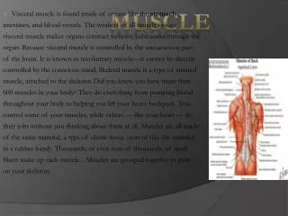
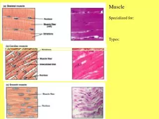
Muscle Types • Skeletal Muscle • large muscle fibers (cells) • striated (banded) • voluntary control • Cardiac Muscle • striated • fibers linked by intercalated disks (electrical synapses) • self-excitation • Smooth Muscle • small tapered fibers • lack striations Figs 17.1, 17.18, and 17.19
Skeletal Muscle Organization • Muscle fibers (cells) • elongate cells, parallel arrangement • sarcolemma – cell membrane • sarcoplamic reticulum (SR) • membraneous internal network • Stores Ca2+ • linked to sarcolemma by transverse tubules Fig 17.1
Skeletal Muscle Organization • myofibrils - intracellular contractile elements • Arranged in sarcomeres (repeated tandem units) • Consist of myofilaments • thick filaments (myosin) • thin filaments (actin) Fig 17.1
Thick Filament Structure • Bundles of several hundred myosin molecules • intertwining tails + globular heads • heads contain • actin binding sites • ATP-hydrolyzing sites • form crossbridges with actin Fig 17.4
Thin Filament Structure • Actin • Primary structural protein • Spherical protein subunits connected in long, double strand • Contains myosin binding site • Tropomyosin • Threadlike proteins • Normally cover myosin binding sites • Troponin • Ca2+ Binding Protein • Holds tropomyosin in place • Ca2+ binding induces shape change that repositions tropomyosin Fig 17.6
Skeletal Muscle Contraction:Sliding Filament Mechanism • Movement of thin filaments over thick • thick filaments are stationary; thin are dragged across thick • sarcomere shortening by increasing overlap of thick and thin filaments Fig 17.3
Crossbridge Cycling • Myosin head binds to actin • Cross bridge bends (Power Stroke) • thin filaments pulled toward center of sarcomere • Cross bridge link broken • Cross bridge ‘unbends’ and binds to next actin molecule Fig 17.5
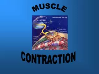
Neural Activation of Skeletal Muscle Contraction • Excitation-Contraction Coupling • Events that link muscle excitation to muscle contraction • Excitation = Action Potential • Brief, rapid depolarization of cell membrane. • Triggers release of Ca2+ into the cytosol of the muscle fiber
Neural Input • Action potential travels down axon to terminal • Exocytosis of acetylcholine (ACh) • ACh diffuses across cleft • ACh opens ACh-gated Na+ channels • Action potential propagates down sarcolemma Fig 17.7
Excitation-Contraction Coupling • T-tubules conduct APs into the cell • Triggers Ca2+ channels in SR to open • Ca2+ released into the cytosol Fig 17.7
Smooth Muscle Contraction • No striations • contractile proteins not arranged in sarcomeres • arranged in fish-net network • allows for extensive contraction, even when stretched Fig 17.18
Smooth Muscle Excitation-Contraction Coupling • Depolarization of sarcolemma opens Ca2+ channels • Ca2+ enters the cell from the extracellular fluid • Ca2+ binds with calmodulin in cytoplasm • Ca2+-calmodulin binds to myosin light chain kinase • activates MLCK • MLCK phosphorylates myosin • needed for myosin to bind actin • Cross-bridge cycling
Whole Muscle Mechanics Types of Contractions • Isometric Contraction • muscle is prevented from shortening • contraction generates force on attachment points • Isotonic Contraction • muscle allowed to shorten upon contraction • muscle moves and object of a given mass Fig 17.9
Isometric Contractions Twitch • response to a single, rapid stimulus Summation • Application of stimuli in rapid succession • Muscle responds to second before fully relaxed from first • Increased tension generated Tetanus • With repeated stimulation at high frequency twitches fuse to form steady tension Fig 17.11
Length-Tension Relationship • Maximum force generated is associated with starting length of muscle • Related to the overlaps of thick and thin filaments • Maximum tension generated at normal in vivo resting length • Too long - pull thick and thin filaments apart • Too short - thin filaments form opposite sides collide Fig 17.12
Isotonic Contraction • Muscle free to shorten with stimulation • shortening distance with load • shortening velocity with load Fig 17.9-17.10
Isotonic Contraction • Contraction Force (N) • Anything that changes the state of motion for an object • Force Cross-Sectional Area • Work (J) • Expresses forces applied to an object to set it in motion • Force × Distance • With load, mass, distance • Max work obtained at ~40% max. load • Work Muscle Mass Fig 17.13
Comparative Muscle Physiology:Vertebrate Skeletal Fiber Types • Fast (Twitch) Fibers • Low myoglobin content (white) • used for rapid movements • large nerve fibers w/ high velocities • anaerobic activity • Slow (Tonic) Fibers • High myoglobin content (red) • used for low-force prolonged contractions • small nerve fibers w/ low velocities • aerobic activity Fig 17.15 Table 17.2
Comparative Muscle Physiology:Fiber Types in Tuna • Tonic Fibers • located in lateral core areas • used for “cruising” • Twitch Fibers • much of the remaining muscle mass • used for short bursts of high activity
Comparative Muscle Physiology:Molluscan Catch Muscle • Used to seal shells of bivalves • Sustained contraction w/o fatigue • Little in O2 consumption • Two groups of neurons innervate muscle • Exciters - releases ACh • causes release of Ca2+ • binds myosin w/o release • Relaxers - releases seratonin • causes release of cAMP • causes Ca2+ release from myosin
Comparative Muscle Physiology:Crustacean Chelipod Muscle • Pinnate (angular) arrangement of fibers • can have more, shorter fibers • 2x increase in force • Few or only single motor unit(s) per muscle • Multiple innervation to each muscle fibers • slow neuron - stimulates tonic activity • fast neuron - stimulates twitch activity • inhibitory neuron - restricts activity Fig 17.17
Comparative Muscle Physiology:Insect Flight Muscle • Synchonous flight muscle • moths, grasshoppers, dragonflies • slow wingbeat frequencies • each contraction is a response to a single nerve impulse • Asynchronous flight muscle • flies, bees mosquitoes • high wingbeat frequencies (100-1000 bps) • too fast for response to individual nerve signals • multiple contractions per nerve signal Box 17.2
Comparative Muscle Physiology:Asynchronous Muscle Function • Fibrillar muscles • attached to walls of thorax • horizontal arrangement • vertical arrangement • not connected to wings • distort shape of thorax Box 17.2
Comparative Muscle Physiology:Asynchronous Muscle Function • upstroke • vertical muscles contract • thorax distorted (click) • stretch horizontal muscles • induced them to contract • downstroke • horizontal muscles contract • distort thorax • stretch vertical muscles • several wingbeats per nerve impulse Box 17.2
Comparative Muscle Physiology:Electric Eels • Electric organs – modified skeletal muscle • Electrocytes – stacked in columns • Respond to signals from motor neurons • All electrocytes in column depolarize spontaneously • Up to 600 V discharge Box 17.1