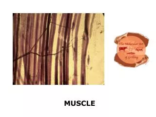
Muscle
Learn about the organization and internal components of skeletal muscle fibers, including sarcomeres, myofibrils, and myofilaments. Explore the functions of skeletal muscles and their vital role in movement, support, and more.


Muscle
E N D
Presentation Transcript
Muscle • Lecture #10 Ch 10 Muscle Muse 11/2/11
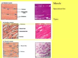
An Introduction to Muscle Tissue • Muscle Tissue • A primary tissue type, divided into • Skeletal muscle • Cardiac muscle • Smooth muscle
Functions of Skeletal Muscles • Produce skeletal movement • Maintain body position • Support soft tissues • Guard openings • Maintain body temperature • Store nutrient reserves
Skeletal Muscle Structures • Muscle tissue (muscle cells or fibers) • Connective tissues • Nerves • Blood vessels
Skeletal Muscle Structures • Organization of Connective Tissues • Muscles have three layers of connective tissues • Epimysium: • exterior collagen layer • connected to deep fascia • Separates muscle from surrounding tissues • Perimysium: (not to be confused with paramecium) • surrounds muscle fiber bundles (fascicles) • contains blood vessel and nerve supply to fascicles • Endomysium: • surrounds individual muscle cells (muscle fibers) • contains capillaries and nerve fibers contacting muscle cells • contains myosatellite cells (stem cells) that repair damage
Skeletal Muscle Structures Figure 10–1 The Organization of Skeletal Muscles.
Epimysium Epimysium Bone Perimysium Endomysium Tendon Muscle fiber in middle of a fascicle (b) Blood vessel Fascicle (wrapped by perimysium) Endomysium (between individual muscle fibers) Perimysium Fascicle Muscle fiber (a) Figure 9.1
Skeletal Muscle Structures • Organization of Connective Tissues • Muscle attachments • Endomysium, perimysium, and epimysium come together: • at ends of muscles • to form connective tissue attachment to bone matrix • i.e.,tendon (bundle) or aponeurosis (sheet)
Skeletal Muscle Structures • Nerves • Skeletal muscles are voluntary muscles, controlled by nerves of the central nervous system (brain and spinal cord) • Blood Vessels • Muscles have extensive vascular systems that • Supply large amounts of oxygen • Supply nutrients • Carry away wastes
Skeletal Muscle: Attachments • Muscles attach: • Directly—epimysium of muscle is fused to the periosteum of bone or perichondrium of cartilage • Indirectly—connective tissue wrappings extend beyond the muscle as a ropelike tendon or sheetlike aponeurosis
Skeletal Muscle Fibers • Are very long • Develop through fusion of mesodermal cells (myoblasts) • Become very large • Contain hundreds of nuclei
Skeletal Muscle Fibers Figure 10–2 The Formation of a Multinucleate Skeletal Muscle Fiber.
Skeletal Muscle Fibers Figure 10–2a The Formation of a Multinucleate Skeletal Muscle Fiber.
Skeletal Muscle Fibers Figure 10–2b The Formation of a Multinucleate Skeletal Muscle Fiber.
Skeletal Muscle Fibers • Internal Organization of Muscle Fibers • The sarcolemma • The cell membrane of a muscle fiber (cell) • Surrounds the sarcoplasm (cytoplasm of muscle fiber) • A change in transmembrane potential begins contractions
Sarcolemma Mitochondrion Myofibril Dark A band Light I band Nucleus (b) Diagram of part of a muscle fiber showing the myofibrils. Onemyofibril is extended afrom the cut end of the fiber.
Skeletal Muscle Fibers • Internal Organization of Muscle Fibers • Transverse tubules (T tubules) • Transmit action potential through cell • Allow entire muscle fiber to contract simultaneously • Have same properties as sarcolemma
Skeletal Muscle Fibers • Internal Organization of Muscle Fibers • Myofibrils • Lengthwise subdivisions within muscle fiber • Made up of bundles of protein filaments (myofilaments) • Myofilaments are responsible for muscle contraction • Types of myofilaments: • thin filaments: • made of the protein actin • thick filaments: • made of the protein myosin
Skeletal Muscle Fibers • Internal Organization of Muscle Fibers • Sarcoplasmic reticulum (SR) • A membranous structure surrounding each myofibril • Helps transmit action potential to myofibril • Similar in structure to smooth endoplasmic reticulum • Forms chambers (terminal cisternae) attached to T tubules
Skeletal Muscle Fibers • Internal Organization of Muscle Fibers • Triad • Is formed by one T tubule and two terminal cisternae • Cisternae: • concentrate Ca2+ (via ion pumps) • release Ca2+ into sarcomeres to begin muscle contraction
Skeletal Muscle Fibers Figure 10–3 The Structure of a Skeletal Muscle Fiber. Show video excitation coupling
Skeletal Muscle Fibers • Internal Organization of Muscle Fibers • Sarcomeres • The contractile units of muscle • Structural units of myofibrils • Form visible patterns within myofibrils • Muscle striations • A striped or striated pattern within myofibrils: • alternating dark, thick filaments (A bands) and light, thin filaments (I bands)
Sarcomere • Smallest contractile unit (functional unit) of a muscle fiber • The region of a myofibril between two successive Z discs • Composed of thick and thin myofilaments made of contractile proteins
Skeletal Muscle Fibers • Internal Organization of Muscle Fibers • Sarcomeres • M Lines and Z Lines: • M line: • the center of the A band • at midline of sarcomere • Z lines: • the centers of the I bands • at two ends of sarcomere
Thin (actin) filament Z disc H zone Z disc Thick (myosin) filament I band A band Sarcomere I band M line (c) Small part of one myofibril enlarged to show the myofilaments responsible for the banding pattern. Each sarcomereextends from one Z disc to the next. Sarcomere Z disc Z disc M line Thin (actin) filament Elastic (titin) filaments Thick (myosin) filament (d) Enlargement of one sarcomere (sectioned lengthwise). Notice the myosin heads on the thick filaments.
Skeletal Muscle Fibers • Internal Organization of Muscle Fibers • Sarcomeres • Zone of overlap: • the densest, darkest area on a light micrograph • where thick and thin filaments overlap • The H Band: • the area around the M line • has thick filaments but no thin filaments
Skeletal Muscle Fibers • Internal Organization of Muscle Fibers • Sarcomeres • Titin: • are strands of protein- quite elastic, like springs • reach from tips of thick filaments to the Z line • stabilize the filaments
Skeletal Muscle Fibers Figure 10–4a Sarcomere Structure.
Skeletal Muscle Fibers Figure 10–4b Sarcomere Structure.
Skeletal Muscle Fibers Figure 10–5 Sarcomere Structure.
Skeletal Muscle Fibers Figure 10–6 Levels of Functional Organization in a Skeletal Muscle.
Skeletal Muscle Fibers Figure 10–6 Levels of Functional Organization in a Skeletal Muscle.
Skeletal Muscle Fibers • Sarcomere Function • Transverse tubules encircle the sarcomere near zones of overlap • Ca2+ released by SR causes thin and thick filaments to interact
Skeletal Muscle Fibers • Muscle Contraction • Is caused by interactions of thick and thin filaments • Structures of protein molecules determine interactions
Skeletal Muscle Fibers • Four Thin Filament Proteins • F-actin (Filamentous actin) • Is two twisted rows of globular G-actin • The active sites on G-actin strands bind to myosin • Nebulin • Holds F-actin strands together • Tropomyosin • Is a double strand • Prevents actin–myosin interaction • Troponin • A globular protein • Binds tropomyosin to G-actin • Controlled by Ca2+
Skeletal Muscle Fibers Figure 10–7a, b Thick and Thin Filaments.
Skeletal Muscle Fibers • Initiating Contraction • Ca2+ binds to receptor on troponin molecule • Troponin–tropomyosin complex changes • Exposes active site of F-actin
Skeletal Muscle Fibers • Thick Filaments • Contain twisted myosin subunits • Contain titin strands that recoil after stretching • The mysosin molecule • Tail: • binds to other myosin molecules • Head: • made of two globular protein subunits • reaches the nearest thin filament
Skeletal Muscle Fibers Figure 10–7c, d Thick and Thin Filaments.
Skeletal Muscle Fibers • Myosin Action • During contraction, myosin heads • Interact with actin filaments, forming cross-bridges • Pivot, producing motion
Skeletal Muscle Fibers • Skeletal Muscle Contraction • Sliding filament theory • Thin filaments of sarcomere slide toward M line, alongside thick filaments • The width of A zone stays the same • Z lines move closer together
Skeletal Muscle Fibers Figure 10–8a Changes in the Appearance of a Sarcomere during the Contraction of a Skeletal Muscle Fiber.
Skeletal Muscle Fibers Figure 10–8b Changes in the Appearance of a Sarcomere during the Contraction of a Skeletal Muscle Fiber.
Skeletal Muscle Fibers • Skeletal Muscle Contraction • The process of contraction • Neural stimulation of sarcolemma: • causes excitation–contraction coupling • Cisternae of SR release Ca2+: • which triggers interaction of thick and thin filaments • consuming ATP and producing tension
The Neuromuscular Junction • Is the location of neural stimulation • Action potential (electrical signal) • Travels along nerve axon • Ends at synaptic terminal • Synaptic terminal: • releases neurotransmitter (acetylcholine or ACh) • into the synaptic cleft (gap between synaptic terminal and motor end plate)
The Neuromuscular Junction Figure 10–10a, b Skeletal Muscle Innervation.
The Neuromuscular Junction Figure 10–10c Skeletal Muscle Innervation.
The Neuromuscular Junction Figure 10–10c Skeletal Muscle Innervation.
The Neuromuscular Junction • The Neurotransmitter • Acetylcholine or ACh • Travels across the synaptic cleft • Binds to membrane receptors on sarcolemma (motor end plate) • Causes sodium–ion rush into sarcoplasm • Is quickly broken down by enzyme (acetylcholinesterase or AChE)
The Neuromuscular Junction Figure 10–10c Skeletal Muscle Innervation.