Muscle
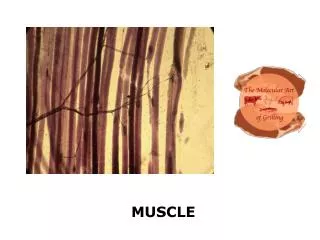
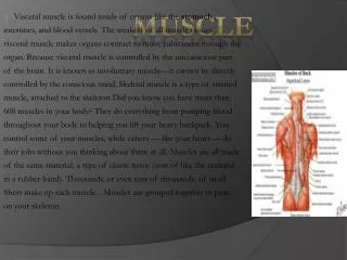
Muscle. Three types of muscle: smooth cardiac skeletal. All muscles require ATP to produce movement. Thus, muscles are chemotransducers. Skeletal Muscle. Muscle organization Muscle innervation Architecture and structure Excitation-contraction Fiber type characteristics


Muscle
E N D
Presentation Transcript
Muscle Three types of muscle: • smooth • cardiac • skeletal All muscles require ATP to produce movement. Thus, muscles are chemotransducers
Skeletal Muscle • Muscle organization • Muscle innervation • Architecture and structure • Excitation-contraction • Fiber type characteristics • Training adaptations • Exam 1 (Feb 8)
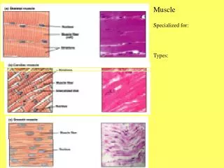
Skeletal muscle organization Connective tissue layers • Epimysium • Perimeysium • Endomysium
Muscle fiber covering • Sarcolemma • basement membrane • plasma membrane • Plasma membrane has • membrane receptors • ion channels • integrins • satellite cells • multinuclei
Muscle Architecture Effect on force output and shortening velocity
Muscle Architecture Muscle architecture
Muscle Architecture Parallel Unipennation Multipennation
Pennation: Effect on Physiological Cross-sectional Area (PCSA) Greater PCSA when fiber is at angle to line of force B A A
Pennation: Effect on Force and Shortening Distance/Velocity Fiber B Fiber A Equal number of sarcomeres in both examples, but Fiber A has longer fiber and smaller PSFA than Fiber B, which allows for greater shortening distance/velocity at sacrifice of force.
Identify which muscles are best suited for force; for speed A B C D
Muscle Architecture • quadriceps and planter flexors designed for force production • larger pennation angles • large PCSAs • hamstrings and dorsiflexors designed for velocity • smaller pennation angles • intermediate PCSAs
Muscle Architecture Summary • Muscles designed to fit purpose of joint • Muscles designed for velocity have longer fiber length and small pennation angle • Muscles designed for force have shorter fiber length and larger pennation angle
Review questions • Describe the difference between a muscle with a fusiform architecture and one with a uni- or multipennate architecture. Identify a muscle for each type of architecture. • Discuss how muscle architecture affects force output and shortening velocity. Provide a general explanation as to why some muscles are designed more for rapid shortening velocity (e.g. hamstrings) or higher force output (e.g. quadriceps muscles).
Muscle Innervation Motoneurons, neuromuscular junctions, motor units
Motoneurons • muscle fibers innervated by large (alpha) myelinated nerves • motoneurons originate from spinal cord • nerve ending ends at neuromuscular junction • motor unit composed of motor neuron and all the fibers it innervates
Action Potential • depolarization – influx of Na+ • repolarization – efflux of K+ • refractory period – hyperpolarization • threshold level – minimal stimulus required to elicit response • muscle and nerve follow “all or nothing principle”
+20 0 -20 -40 -60 -80 Membrane potential (mV) Time (ms) K+ K+ K+ Na+ Na+ K+ Na+ Na+ Na+ Na+ Na+ Na+ Na+ Na+ Na+ Na+ Na+ Na+ Na+ Na+ channel Na+-K+ exchange pump K+ channel ATPase K+ K+ K+ K+ K+ K+ K+ K+ K+ K+ ADP K+ K+ K+ K+ K+ K+ Na+ Pi Na+ Na+ Na+ intracellular ATP
Electromyography (EMG) Describe the relative weights being lifted
Review questions • Define the motor unit. • Describe the events that occur as an action potential approaches the nerve terminal. • Explain the purpose of acetylcholinesterase and the consequences of its absence. • A common agent found in flea powders is a low dose of an antiacetylcholinesterase inhibitor. Explain the effects that the flea powder would have on fleas. • Explain the interpretation of an EMG tracing.
Skeletal Muscle Structure • sarcomeres (smallest functional unit) are linked end-to-end to form myofibrils • myofibrils are bunched to form fibers • sarcomeres are composed of thick and thin filaments
Scanning EM 1 4 2 5 3
Thick Filament • composed of numerous myosin protein strands • flexible “heads” protrude outward all around filament (except center) • myosin heads attach to “active” sites on actin (thin) filament • myosin heads contain ATPase to break down ATP
Thin Filament • actin - two protein strands twisted around each other, contain “active sites” • tropomyosin - thin strand laying in actin groove that covers active sites • troponin - attached to actin and tropomyosin strands; has strong affinity for Ca2+ Composed of three proteins
Cytoskeleton (structural) proteins • M-band– located in middle of thick filament; provides structural support to myosin filaments; contains creatine kinase (CK) • Titan–connects myosin filament to Z-disk; stabilizes myosin in middle of sarcomere. • Z-disk –thin filaments attachment; composed of several cytoskeletal proteins
Transverse Tubule • in human skeletal muscle, each sarcomere has two transverse tubules running perpendicular to fiber • T-tubules extend through fiber and have openings at sarcolemma allowing communication with plasma • cardiac fibers have only one T-tubule which lies at Z-line
Sarcoplasmic Reticulum (SR) • made up of terminal cisternae and longitudinal tubules • serves as a storage depot for Ca2+ • terminal cisternae abut T-tubules • longitudinal tubules cover myofibrils and connect terminal cisternae
1. On what component does Ca2+ bind to? • Sarcoplasmic reticulum • Myosin heads • Troponin • Tropomyosin • 2. What protein returns Ca2+ to the sarcoplasmic reticulum? • Myosin head • Ca2+ pump • Ca2+ channels • tropomyosin
Review questions • Describe the myosin filament of a skeletal muscle fiber. Include a detailed description and function of the myosin head. • Describe the thin filament of a skeletal muscle fiber. • Describe the cytoskeleton proteins and their functions in the sarcomere. • Describe the sarcoplasmic reticulum and its role in excitation-contraction.
Excitation-Contraction How muscle contracts
Excitation-Contraction Coupling • action potentials, generated at neuromuscular junction travel around sarcolemma and through T-tubules • T-tubules signal SR to release Ca2+ into sarcoplasm (cytosol) • Ca2+ saturates troponin (in non-fatigued state) • troponin undergoes conformational change that lifts tropomyosin away from actin filament
E-C Coupling (cont.) • myosin head attaches to active site on actin filament • after attaching to actin, myosin head moves actin-myosin complex forward and releases ADP and Pi • ATP binds with myosin head, which releases actin, and returns to original position • in resting state, myosin head contains partially hydrolyzed ATP (ADP and Pi)
E-C Coupling (cont.) • entire cycle takes ~50 ms although myosin heads are attached for ~2 ms • a single cross-bridge produces 3-4 pN and shortens 10 nm • as long as action potentials continue, Ca2+ will continue to be released • when action potentials cease, SR Ca2+ pumps return Ca2+ ceasing contractions • skeletal motor units follow “all or nothing” principle
Excitation-Contraction • AP causes vesicles to release Ach • Muscle AP travels down t-tubules • SR releases Ca2+ into sarcoplasm • Ca2+ binds to troponin • Myosin heads bind to actin; mysoin ATPase splits ATP • ATP binds to myosin heat; releases from actin • Crossbridge action continues while Ca2+ is present • When AP stops, Ca2+ pumped back to SR • Tropomyosin covers active sites
EC Coupling • QuickTime Movie of sliding filaments • http://www.sci.sdsu.edu/movies/actin_myosin.html • Click on Link • Click on Actin Myosin Crossbridge 3D Animation
3. What will happen if ATP is depleted in muscle? • Nothing • Muscle will relax • Muscle will not relax 4. What will happen if sarcoplasmic reticulum of fiber is enhanced? • Fiber will develop tension more quickly • Fiber will relax more quickly • Nothing • Both a and b will occur