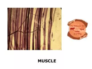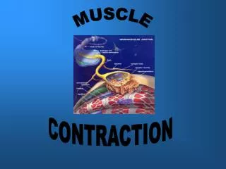
MUSCLE
MUSCLE. PHYSIOLOGY. MUSCLE. Muscle is an organ: several tissues working together Muscle Nerve Blood Fibrous connective Three types of muscle; the description of muscle function will deal with skeletal. MUSCLE TYPES. SKELETAL

MUSCLE
E N D
Presentation Transcript
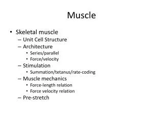
MUSCLE PHYSIOLOGY
MUSCLE • Muscle is an organ: several tissues working together • Muscle • Nerve • Blood • Fibrous connective • Three types of muscle; the description of muscle function will deal with skeletal
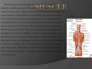
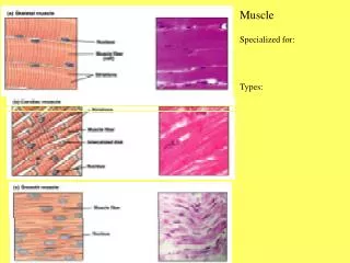
MUSCLE TYPES • SKELETAL • Covers bones; responsible for voluntary movements • striated • SMOOTH • Intestinal tract, etc. involuntary • CARDIAC • Found only in the heart
FASCIA • Fibrous connective in layers • Separates adjacent muscles • Holds position • Surrounds each muscle and extends into tendon to intertwine with periosteum • Part of tendon
APONEUROSIS • Broad fibrous sheet • Connect muscle to muscle • Found in lower back and on skull
MYSIUMS • EPIMYSIUM • Around muscle itself (close) • PERIMYSIUM • Extends in from epimysium • Separates muscle into bundles called FASCICULI • ENDOMYSIUM • Thin layer around each muscle fiber (cell)
MUSCLE • P. 178; FIGURE 8.1 • Tan box p. 179 • MYOFIBER = MUSCLE CELL = FIBER
MYOFIBER • Muscle cell • Contracts when stimulated, then relaxes • Thin, elongated, round ends and may extend the full length of the muscle • Cell membrane is SARCOLEMMA • Cytoplasm is SARCOPLASM • Many small, oval nuclei and mitochondria
MYOFIBER • Many thread-like MYOFIBRILS that run parallel • MYOFIBRILS are made of MYOFILAMENTS • 2 types of filaments • ACTIN: light, thinner • MYOSIN: dark, thicker
MYOFIBER • Striations caused by myosin and actin • Diagram p. 179 • Z lines (ends of sarcomere, runs through middle of actin) • A band ( dark, actin and myosin) • I band (light, actin) • SARCOMERE is one Z line to the next
SARCOMERE • Z line to Z line • SARCOPLASMIC RETICULUM • Like endoplasmic reticulum • CISTERNAE • Enlarged portions of SR; extend around myofibril • T-TUBULES (transverse) • Run transversely
NEUROMUSCULAR JUNCTION T-tubules and reticulum activate muscle contraction when stimulated • NEUROMUSCULAR JUNCTION • Motor neuron: nerve cell to muscle cell • Motor end plate: on muscle fiber • Neurotransmittors: acetylcholine (ATCh)
NEUROMUSCULAR JUNCTION • MOTOR UNIT is the motor neuron and muscle fiber or fibers working together • Organized fibers controlled by a single motor neuron
MUSCLE PROTEINS • 1) ACTIN • Thin • Globular with binding sites to accept myosin and form cross bridges • Many actin molecules twist into double strand (helix)to form filament
MUSCLE PROTEINS • 2) MYOSIN • 2 twisted protein strands with extending cross bridges • Thick Actin and myosin slide past each other and shorten the muscle
MUSCLE PROTEINS • 3) TROPONIN • Work to expose binding sites • Located on actin • 4) TROPOMYOSIN • Work to expose binding site • On actin P. 182
SLIDING FILAMENT THEORY • The heads of the myosin crossbridges attach to actin binding sites and bend slightly, pulling the actin fibers with it. • Head releases and attaches immediately to the next actin-binding site • Hand-over-hand pulling idea (sailing) • ATPase enzyme on head of myosin releases PHOSPHATE from ATP; this energy cocks myosin head to prepare to attach to actin • P. 183-184
CONTRACTION • ACETYLCHOLINE • Neurotransmitter • Synthesized in cytoplasm of nerve • Stored in vesicles • When impulse reaches the end of the motor neuron, ATCh is released into synaptic cleft between neuron and motor end plate of muscle • P. 181
CONTRACTION • ATCh rapidly diffuses combines with receptor proteins of muscle cell membrane impulse passes in all directions over surface of muscle fiber membrane (sarcolemma) and through T-tubules to reticulum • Sarcoplasmic reticulum has high Ca++ cisternae become more permeable to these ions and they spread into the sarcoplasm • P. 181
CONTRACTION • When Calcium is high troponin and tropomysium interact to expose binding sites on actin linkage forms and muscle contracts • Acetylcholinterase is the enzyme that decomposes ATCh right after release to prevent continued response • Calcium transported back to reticulum
TABLE P. 184 • Tan box P. 184 BOTULISM • Clostridium botulism produces a toxin that prevents the release of ACTh • Causes botulism: paralysis of nerves (esp. those involved in breathing)
ENERGY SOURCES • ATP • Only a short supply in fiber • Need to regenerate ATP (remember ADP + P ATP) • CREATINE PHOSPHATE • Has high energy bonds and 4-6 times more abundant in fibers • BUT can’t be use directly
ENERGY SOURCES • CREATINE PHOSPHATE • Stores excess energy released from mitochondria • When ATP insufficient, creatine phosphokinase (enzyme) causes creatine phosphate to be formed • As ATP decomposes, energy from creatine phosphate is transferred to ADP converting it back to ATP
ENERGY SOURCES • Active muscle rapidly uses CP • When it is gone, the muscle relies on glucose from cellular respiration • Resting muscle has reserves of: • ATP • Glycogen • CP • Ca++
OXYGEN SUPPLY • GLYCOLYSIS (no O2); the rest of respiration needs oxygen • Oxygen carried by RBC • Muscle has MYOGLOBIN pigment and can carry some oxygen (red-brown color of muscle)
OXYGEN SUPPLY • Continued contraction depletes oxygen • Now muscle depends on anaerobic respiration • Makes pyruvic acid • With continued low oxygen this becomes LACTIC ACID and diffuses into blood and liver (stinging sensation) • Addition of oxygen converts lactic acid back to glucose
STRENUOUS EXERCISE • Lactic acid accumulates • OXYGEN DEBT is the amount of oxygen the liver cells require to convert accumulated lactic acid back to glucose • Takes several hours to complete
STRENUOUS EXERCISE • After strenuous exercise lactic acid (70%) that is converted back to glucose goes back to muscle to build glycogen supply
MUSCLE FATIGUE • Strenuous exercise over a prolonged period • Muscle loses ability to contract • Caused by: • Interruption of blood supply • Lack of ATCh • Accumulation of lactic acid which LOWERS the pH and muscles no longer contract ****
RIGOR MORTIS • Tan box p. • Several hours after death • Continues for > 72 hrs • Caused by increase in membrane permeability to calcium and decrease in ATP • Prevents relaxation; actin and myosin remain linked until the muscle decomposes
RESPONSES • Heat production • Most of the heat produced goes to help maintain body temperature (60%) • Threshold stimulus • A single fiber can respond • Stimulus has to reach a certain strength to cause contraction
RESPONSES • ALL-OR-NOTHING • A fiber exposed to threshold stimuli will respond fully • Increased stimuli does NOT increase the degree of contraction • Fiber normally does NOT contract partially • THE NUMBER OF FIBERS STIMULATED IS IMPORTANT!!!
STEROIDS • P. 187 • HAND-OUT
RECORDING CONTRACTION • MYOGRAM • Record of contraction pattern • TWITCH • Single contraction that lasts a fraction of a second • LATENT PERIOD • Delay at the start 0.01 sec. • LATENT PERIOD PERIOD OF CONTRACTION RELAXATION PERIOD
SUSTAINED CONTRACTION • SUMMATION • When exposed to a series of stimuli of increasing frequency, the muscle does not completely relax; individual twitches combine SUMMATION • Holds contraction (sustain) • RECRUITMENT • Nearby motor units respond, each at its own threshold • Remember: a motor unit is organized fibers controlled by one neuron
SUSTAINED CONTRACTION • Twitches combine, strength of contraction may increase • SUMMATION + RECRUITMENT = SUSTAINED CONTRACTION • Needed for walking, lifting, etc. Tetanic contraction is a forceful, sustained contraction with NO relaxation (power lift)