Advanced Surface Analysis Techniques: SIMS, AES, LEED & RHEED
340 likes | 705 Vues
Explore advanced surface analysis techniques, including SIMS, AES, LEED, and RHEED. Learn about their principles, advantages, and disadvantages, and how they are used for chemical and structural analysis. Discover the capabilities and limitations of these sophisticated tools in materials science research.

Advanced Surface Analysis Techniques: SIMS, AES, LEED & RHEED
E N D
Presentation Transcript
Surface analysis techniques part I Yaniv Rosen
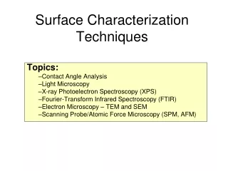
Surface Analysis Techniques • Chemical Analysis • SIMS (Secondary ion mass spectroscopy) • AES (Auger electron spectroscopy) • Structural Analysis • LEED (Low-energy electron diffraction) • RHEED (Reflection high energy electron diffraction)
Secondary ion mass spectroscopy • Sputter sample with high energy ions for example 4KeV Ar . • Surface material is released. • Use conventional ion mass spectrometers to determine composition. +
Advantages of SIMS • Very sensitive – can reach parts per billion range. • Ability to do depth profiling.
Disadvantages of SIMS • Only ionized particles are measured.
Disadvantages of SIMS • Only ionized particles are measured. • Sputtering not necessarily even. • Different levels of ionization.
Disadvantages of SIMS • Only ionized particles are measured. • Sputtering not necessarily even. • Different levels of ionization. • Intrinsically destructive: • Dynamic SIMS • 1nA/cm² • 1µm/hr • Static SIMS • 1mA/cm² • 1Å/hr
Auger Electron Spectroscopy • Fire ~100eV-5keV electrons at sample • Electron knocked out of atomic core • Higher level electron falls into hole. • Outer shell electron emitted with excess energy. • Measure energy of emitted electron: KE=Ea-Eb-Ec* Ea Eb Ec
Why use AES? • Easy to detect 1% impurity in monolayer. • Beam of electrons can be focused and moved easily – provides high resolution. • Image can be compared simultaneously with SEM (Scanning electron microscope) image. • Good transition rates for smaller elements – can get signal for Li.
AES disadvantages • High resolution and fast rates can cause sample damage. • Theoretical predictions are complicated.
AES disadvantages • High resolution and fast rates can cause sample damage. • Theoretical predictions are complicated. • Absolute quantification not attempted
Low-energy electron diffraction • Fire 20-300eV electrons at sample in Ultra-High Vacuum (UHV~10^-9 torr) • Diffraction and elastic scattering occurs • Accelerate electrons towards florescent mesh. • Pattern should match reciprocal lattice.
LEED Advantages • Small mean-free path through the material • Same instrumentation as AES – can be placed in same apparatus. • Averages over small defects in the periodicity.
LEED Disadvantages • Adsorbates change the configuration. • Possible to have multiple configurations from one spectra. • Difficult theory when more then one atom in base cell.
Reflection high energy electron diffraction • 30-100KeV. • Fire high energy electrons at a shallow angle. • Use phosphorus screen to detect diffraction pattern.
RHEED Advantages • Electrons have high energy so they do not need help reacting with phosphorus. • Sensitive to local defects – used in MBEs (Molecular-beam epitaxy) systems to grow semi-conductors.
RHEED Disadvantages • Mostly concerned with qualitative descriptions of surface as opposed to quantitative diffracted beam intensity. • Needs high vacuum so electrons are not deflected.
Conclusion • Chemical Analysis • SIMS – Destructive but very sensitive to impurities. • AES – high resolution for chemical analysis. Uses same instrumentation as LEED. • Structural Analysis • LEED – Bulk structure analysis. • RHEED – Detects local defects.
Principle • X-ray (or UV) photons excite electrons to continuum states. • Electron kinetic energy (eKE) related to binding energy of the initial state by: eKE = hv – BE – δE eKE E=0 hv BE Core Level
Method Energy Analyzer Electrostatic Lens Detector X-rays/ UV Photoelectrons Sample Huefner et. al. 1996
Typical Properties • Resolution: • XPS ~.25eV (100 - >1000eV) • UPS ~.10eV (10 - 40eV) • Detection limits of 1 part in 10k to 100k for long measurements • Can sample first ~10nm
Limitations • Surface properties interfere with attempts to measure bulk properties • Sample degradation • Charge loss • Radiation damage • Lower and upper bounds on analysis spot size (micron-mm) • UHV requirement, no magnetic field, low electric field
Chemical Shift • PES lines affected by surroundings Neudachina et. al. 2005 Uhrberg et. al. 1998
Ad/Desorption Properties Characterize potential energy surface of final ionized state adsorbed onto substrate AB+ + S (A+B+) + S AB + S hv (A+B) + S Fohlisch et. al. 1998
ARPES Conservation laws eKE = hv – BE pef = phv + pei 0 θ Damascelli 2004
Valence Band Characterization Damascelli 2004
Inverse XPS Study unoccupied bands • Provides complimentary information to photoemission • Directly measure density of states above EF • Can use analogous techniques to PES Smith 1988