Download
1 / 38
400 likes | 576 Vues
Learn about the importance of flexibility in restoring range of motion post-injury. Explore factors affecting flexibility, soft tissue properties, and muscle anatomy that impact mobility. Discover active and passive range of motion techniques for successful recovery.

E N D
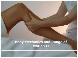
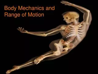
Importance of Flexibility • Important Goal: Restore or improve to normal pre-injury range of motion • With injury there is generally some degree of lost range of motion • Due to pain, swelling, muscle guarding, &/or inactivity resulting in tissue shortening • Need to encourage stretching exercises • Restricted range of motion can impact performance & result in uncoordinated motion • Essential for successful physical performance & injury prevention
Flexibility • Ability of neuromuscular system to allow for efficient movement of a joint or series of joint through a full, non-restricted pain free range of motion
Anatomic Factors Impacting Flexibility • Muscles • Increasing flexibility relies on the elastic properties of muscle • Length can be changed over time • Connective Tissue • Ligaments & joint capsules, while possessing some elastic properties, can lose their elasticity during periods of disuse & immobilization • Bony Structures • Can limit end point range • Bony prominences can also stop movements at normal end points in the range • Fat • Can act as a wedge between lever arms • Restricts movement wherever it is found
Skin • Injury or surgical procedure may alter skin – variable in elasticity • Skin adheres to underlying tissue • Neural tissue • Develops tightness as a result of compression, chronic repetitive microtrauma, muscle imbalances, joint dysfunction or morphological adaptations due to posture • Could stimulate nociceptors & pain • Cause muscle guarding & spasm to protect irritated neural structures • Neural fibrosis ultimately results causing decreased elasticity & restricted motion • Except for bone structure, age & gender all other flexibility limiting factors can be modified & altered to increase range of motion
Soft Tissue Properties that Affect Immobilization & Elongation • Responses that affect soft tissue during stretching • Velocity, intensity, frequency & duration of stretch force • Temperature of tissues • Elasticity – ability of soft tissue to return to its resting length after passive stretch • Plasticity– tendency of soft tissue to assume a new & greater length after stretch force has been removed
Soft Tissue Properties that Affect Immobilization & Elongation • Contractile tissue: gives muscle characteristics of contractility & irritability • Noncontractile tissue: has same properties as all CT, including ability to resist deforming forces as well as viscoelasticity • CT structures of muscle-tendon unit • Epimysium – enveloping fascial sheath • Perimysium – encases bundles of fasciculi • Endomysium – innermost layer that separates individual m. fibers & myofibrils
Muscle Anatomy • Made up of many muscle fibers that lie parallel with one another • Single fiber – made up of many myofibrils • Myofibrils - composed of sarcomeres • Sarcomere – contractile unit of the myofibril • Gives muscle ability to contract & relax • Composed of overlapping myofilaments of Actin & Myosin (form cross-bridges) • Motor unit stimulated = m. contraction -actin-myosin filaments slide together & the muscle actively shortens • Muscle relaxes = cross-bridges slide apart slightly & the muscle returns to its resting length
Myofilament • Interlocking Mesh Structure • A myofilament shows several distinct bands • Each band has been given a special letter • The lightest (least electron dense) band is the “I band” • Consists primarily of actin • In the center of the “I band” is the “Z-line”, an electron dense line • The wide, dark band is the “A band” • Consists primarily of myosin • In the middle of the “A band” is the “M line”, another dense line
Noncontractile Tissue • Made up of: • Collagen – resist tensile deformation & are responsible for strength & stiffness of tissue, elongates quickly under light loads • Elastin - extensibility • Reticulin fibers – bulk • Ground substance – proteoglycans (PGs) & glycoproteins; • PGs hydrate matrix, stabilize collagen network, resist compressive forces; • Glycoproteins provide linkage between matrix components & between cells & matrix opponents • Mechanical behavior is determined by proportion of collagen & elastin fibers & structural orientation of the fibers • High collagen, low PGs – resist high tensile loads • High collagen content tissue = greater stability (tendons)
Active & Passive Range of Motion • Active range of motion (AROM) • Dynamic flexibility • Joint movement via muscle contraction • Ability to move a joint with little resistance • Passive range of motion (PROM) • Static flexibility • Motion of joint to end points without muscle contraction • Critical in injury prevention • Muscles can be forced to stretch beyond “normal” limits • Without elasticity it is likely that the musculotendinous unit will be injured • During athletic activity • Must be able to move through unrestricted range • Must have elasticity for additional stretch encountered during activity
Measuring Range of Motion • Essential to assess improvement during rehabilitation • Goniometer • Utilizes alignment of two arms parallel to longitudinal axis of two segments involved in motion • Relatively accurate tool • Ensures accuracy standardize techniques & methods of recording AROM & PROM
Agonist vs. Antagonist Muscles • Joints are capable of multiple movements • Example: • Quadriceps will extend knee with contraction • Quads (muscle producing movement) = agonist • Hamstrings will stretch during knee extension • Hamstrings undergoing stretch = antagonist • Agonist & antagonist work together to produce smooth coordinated movements • Muscles that work together function synergistically • What is another pair of agonist/antagonist muscles?
Stretching Techniques • Ballistic • Bouncing movement in which repetitive contractions of agonist work to stretch antagonist muscle • Static stretching • Stretch to point of discomfort & holding at that point for period of time • Proprioceptive Neuromuscular Facilitation (PNF) • Involves alternating contractions & stretches • Myofascial & neural tissue stretching • Enhances neuromuscular system’s ability to control movement
Ballistic Stretching • Need to be careful when performing this stretch • Possible soreness due to uncontrolled forces within muscle created by bouncing • May result in tissue damage • Should be incorporated into a program to allow body to adapt & reduce likelihood of injury • Incorporate into later stages of rehabilitation
Static Stretching • Passively stretching given antagonist • 6-8 second hold in maximal position of stretch • Go to point of discomfort & back off slightly • Hold for 15-30 seconds (do this 3-4 times) • Can be accomplished utilizing agonist • Controlled movement, less chance of injury
Proprioceptive Neuromuscular Facilitation • Three techniques that combine alternating isometric or isotonic contractions & relaxation of both agonist & antagonists • Slow-reversal-hold-relax • Contract-relax • Hold-relax • Hold Relax (HR) • Isometric contraction of antagonist followed by concentric contraction of agonist with light pressure • Facilitates stretch of antagonist • Effective with muscle tension on one side of joint
Contract Relax (CR) • Moves body passively into agonist pattern • Athlete instructed to contract antagonist isotonically against resistance • Athlete then relaxes & allow athletic trainer to push body further (passively) into agonist pattern • Utilized when flexibility is limited due to muscle tightness • Slow Reversal-Hold-Relax (SRHR) • Isotonic contraction of agonist • Follow with isometric contraction of antagonist • During relax phase antagonist is relaxed while agonist contracts in agonist pattern • Results in stretch of antagonist • Useful to stretch antagonist
Comparing Stretching Techniques • Ballistic stretching is recommended for athletes engaged in dynamic activity • Static stretching most widely used • Safe & effective • PNF techniques • Capable of producing dramatic increases in ROM • Limitation – partner is required • Maintaining flexibility • Can decrease considerable after only 2 weeks • Should be engaged in at least once per week
Stretching Neural Structures • Requires differentiation between musculotendinous & neural tightness • Assess movements that create tension in neural structures • May cause numbness & tingling • Straight-leg raise example
Myofascial Release Stretching • Techniques used to relieve abnormally tight fascia • Myofascial restrictions are unpredictable & may occur in different planes & directions • Requires specialized training & in depth understanding of fascial system • Fascia • Connective tissue that runs throughout the body & establishes interconnectedness of body • If altered or injured can result in localized response at focal point of injury or away from injury site • Responds to gentle pressure
Sometimes called: Soft-tissue Mobilization • Treatment • Localize restriction • Considerably more subjective component & relies heavily on clinician’s experience • Focuses on large treatment area • Work superficial to deep • Joint mobilizations may follow • Tissue stretching & elongation as well as strengthening should follow • Postural re-training may also be required • Dramatic results may occur • Treatment should be done at least 3 times per week • Perform manually or via foam roller
Neurophysiological Basis of Stretching Stretch Reflex • Muscle is placed on stretch – muscle spindle • Muscle spindles fire relaying info. to spinal cord • Spinal cord relays message to golgi tendon & increases tension • After 6 seconds, golgi tendon organ (GTO) relays signal for muscle tension to decrease • Cause reflex relaxation • Prevents injury - protective mechanism • Ballistic stretching does not allow this overriding response by GTO
With static stretching GTO’s are able to override impulses from muscle spindle following initial reflex resistance • Allows muscle to remain stretched without injury • PNF benefits greatly from these principles • With slow-reversal hold technique, maximal contraction of muscle stimulates GTO reflex relaxation before stretch applied
Autogenic inhibition • Relaxation of antagonist during contraction • During relaxation phase, antagonist is placed under stretch but assisted by agonist contraction to pull further into stretch • GTO is protective mechanism that inhibits tension in the muscle • Reciprocal inhibition • Isotonic contraction of an agonist muscle elicits a reflex relaxation of antagonist muscle group - (protect against injury)
Effect of Stretching on Physical & Mechanical Properties of Muscle • Physical lengthening of muscle occurs due to reflex relaxation • Contractile & non-contractile elements of muscle dictate capability of deformation & recovery • Both resist deformation • Deformation is dependent on degree of stretch & velocity • Non-contractile – limit degree • Contractile – limit velocity • Greater stretch = more non-contractile components contribute
Stretches sustained long enough (autogenic inhibition) result in viscoelastic & plastic changes in collagen & elastin • Viscoelastic changes allow slow deformation & imperfect recovery (not permanent) • Plastic changes result in permanent changes in length • Greater velocity = greater chance for exceeding tissue capacity (viscoelastic & plastic)
Effects of Stretching On Kinetic Chain • Joint hypomobility causes: • Faulty posture • Muscular imbalance • Abnormal neuromuscular control • Alteration in arthrokinematics • Change in muscle tension to reduce translation • Alters degrees of tension & activation in synergist, stabilizers & neutralizers • Compensatory response
Impact on length-tension relationships Alters force couples & arthrokinematics Impacts normal force couple relationships & creates kinetic chain reaction Impacts synergistic function of kinetic chain Causes abnormal joint & tissue stresses, neural compromise & vascular/lymphatic stasis Alters recruitment strategies & stabilization Alters neuromuscular efficiency impacting activation/firing sequence Additionally altered joint function & stress response Can causes reciprocal inhibition Increases muscle spindle activity May impart inhibitory response (decreased neuromuscular control) Result = synergistic dominance – synergist compensatory action for weak & inhibited muscle Muscle Tightness & Hypertonicity
Importance of Warm-up Prior to Stretching • Intramuscular temperature should be increased prior to stretching • Positive effect on ability of collagen & elastin to deform • Enhances reflexive relaxation associated with golgi tendon organs • Optimal temperature 39oC/103oF • To increase = low intensity, warm-up type exercise or modalities • Exercise should be primary means of warm-up • Environment - Heat vs. Cold
Flexibility vs. Strength • Co-exist • Muscle bound • Negative connotation • Loss of motion • Encourage full pain free movements during rehabilitation • Strength training will provide individual with ability to develop dynamic flexibility through full range of motion • Develop more powerful & coordinated movements
Warm-up Overload or stretch beyond normal range Not to point of pain Stretch to point of resistance Increases in range will be specific to muscle being stretched Use caution when stretching around painful joints Avoid overstretching ligaments & capsules Exercise caution with low back & neck stretches Stretch from seated position to reduce stress on back Continue normal breathing while stretching For improvements in ROM, utilize static & PNF stretching techniques Ballistic stretching should be used by those who possess flexibility & are accustomed to it Ballistic stretching should follow period of static stretching Stretching should be performed a minimum of 3 times per week For maximum gains stretching 5-6 times per week is ideal Guidelines & Precautions for Stretching