Download
1 / 61
640 likes | 855 Vues
Explore the gross anatomy of lower extremity veins, examining the venous system from legs to heart. Learn about paired veins, femoral and common femoral veins, the role of perforators, and factors affecting venous flow. Discover the importance of the Greater Saphenous Vein. Enhance your knowledge for comprehensive sonography.

E N D
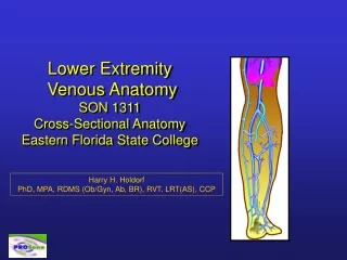
Lower Extremity Venous AnatomySON 1311Cross-Sectional AnatomyEastern Florida State College Harry H. Holdorf PhD, MPA, RDMS (Ob/Gyn, Ab, BR), RVT, LRT(AS), CCP
Cross-Sectional Anatomy • Venous Legs • LE Venous system Q: Why are we covering the LE venous system so early in our education? A: The LE venous system is more often than not, a straight line. If the beginning sonographer can master the LE venous system, all the other studies may be easier to grasp.
Venous Gross Anatomy • Lower Extremity Veins • Note: The anatomical relationship of the veins to the heart is the same as for the arteries. Veins located at the ankle are considered distal: while veins located closer to the heart (e.g., Femoral) are considered more proximal • Note: Be sure to know the orientation of vessels from medial to lateral and from lateral to medial
The paired, deep veins of the calf (Anterior Tibials, Peroneals, and posterior tibilas, follow the corresponding arteries: are called comitantes (corresponding veins).
Paired peroneal veins (PerV) • Formed by confluence of venules • Empties the lateral leg • Paired veins may form common trunk and carry blood cephalad into tibial-peroneal trunk • Paired posterior tibial veins (PTV) • Formed by confluence of venules • Empties back of leg • Paired veins may form common trunk and carry blood cephalad into tibial-peroneal trunk
Paired anterior tibial veins (ATV) • Formed by confluence of venules • Empties front of leg • Popliteal vein (PopV) • Formed by union of ATV and Tib-Peroneal Trunk • Usually just below the knee • Becomes femoral vein (previously called superficial femoral vein) when passes through adductor hiatus in lower thigh
Adductor hiatus The adductor hiatus is a hiatus (gap) between the adductor magnus muscle and the femur that allows the passage of the femoral vessels from the anterior thigh to the posterior thigh and then the popliteal fossa.
Femoral Vein (FV) • Popliteal vein becomes FV when vein passed through adductor hiatus • Common Femoral Vein (CFV) • Formed by joining of FV & Deep femoral vein
External iliac Vein (EIV) • Common femoral vein becomes EIV when passes through inguinal ligament • Common Iliac Vein (CIV) • Formed by confluence of external and internal iliac veins
Because the left common iliac vein passes under the right common iliac artery, extrinsic compression may be evident. • This pressure point may account for left sided DVT; also known as May-Thurner Syndrome
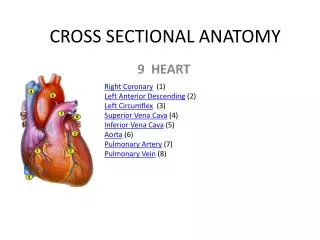
Inferior Vena Cava (IVC) • Formed by confluence of common iliac veins • Commonly at level of 5th lumbar vertebra • Carries blood into right atrium of heart
Superficial Veins • Small saphenous (formally lesser) vein (SSV) • Ascends back of calf joining popliteal vein
Superficial Veins • Great (formerly Greater) Saphenous Vein (GSV) • Longest vein in the body, originating on dorsum of foot, traveling medially to saphenofemoral junction in the groin (about level of CFA bifurcation)
Perforators • Carry blood from superficial veins into deep veins • Posterior arch vein • Has three ankle perforators • Plays major role in development of venous stasis ulcers
Blood Flows from superficial veins (S) to the deep venous system (D)
Factors Affecting Venous Flow • Venous-Skeletal muscle pump ‘Venous heart’ • The GREAT MUSCLE PUMP = Calf muscle • Muscle contraction squeezes vein propelling blood upward-forward
Effective Calf Muscle Pump • Blood moves from superficial system (s) to deep system (D) • Competent valves prevent reflux • Venous volume and pressure decreases • Venous return to heart increases
Ineffective Calf Muscle Pump • Incompetent valves cause reflux • Venous volume and pressure increases • Results in venous pooling and ambulatory venous hypertension.
RespirationInspiration (Phasic) • Decrease in intra-thoracic pressure • Increases blood flow from upper extremities • Increase in intra-abdominal pressure • Decreases blood flow from lower extremities
RespirationExhalation • Increase in intra-thoracic pressure • Decreases blood flow from upper extremities • Decrease in intra-abdominal pressure • Increases blood from lower extremities
Valsalva Maneuver Patient takes in deep breath & holds it, then bears down (as if having a bowel movement) Intra-thoracic and intra-abdominal pressure increases significantly All venous return halted Veins will enlarge
Importance of the Greater Saphenous Vein • The great saphenous vein (GSV), previously also called the long saphenous vein, is a large, subcutaneous, superficial vein of the leg. It is the longest vein in the body running along the medial length of the leg. Clinical significance • Pathology of the great saphenous vein is relatively common, but in isolation typically not life-threatening. • Varicose veins: The great saphenous vein, like other superficial veins, can become varicose; swollen, twisted and lengthened, and generally considered to be unsightly. • Thrombophlebitis The GSV can thrombose. This type of phlebitis of the GSV is usually not life-threatening in isolation; however, if the blood clot is located near the sapheno-femoral junction or near a perforator vein, a clot fragment can migrate to the deep venous system and to the pulmonary circulation. Also it can be associated with, or progress to a deep vein thrombosis which must be treated. The vein is often removed by cardiac surgeons and used for auto transplantation in coronary artery bypass operations, when arterial grafts are not available or many grafts are required, such as in a triple bypass or quadruple bypass.
Vein Mapping for Coronary Artery bypass graft surgery • A tourniquet is placed around the upper thigh or the patient is placed in reverse Trendelenberg to aid in venous filling. • The LSV is carefully traced onto the exterior of the leg using a surgical marker. Beginning distally at the medial malleolus, a generous amount of ultrasound gel is applied to the donor leg. • The focus is set to a depth of 2.8 cm to the scan area below the knee and may be adjusted to greater depths as the thigh is reached. • The vein is identified and confirmed on the ultrasound screen as a tubular, compressible structure. • The LSV is accurately followed up the medial aspect of the leg and marked for the entire length of its course. • The caliber of the vein is assessed and measured at multiple sites and adjustment is made for distension.
Superficial Veins - GSV Images courtesy of Phillips - ATL
Superficial Veins - GSV Sheath Greater saphenous sheath
Duplex-Color Flow Imaging • Identify venous thrombosis – help differentiate acute from chronic • Evaluate non-occluding/partial thrombus • Detect calf lesions • Distinguish between extrinsic compression and intrinsic obstruction • Evaluate soft tissue masses • Detect venous incompetence • Document re-canalized channels or collateralization
Limitations of Duplex Imaging • Sources of false-Positive studies include: • Extrinsic compression: e.g., tumors such as SVC syndrome, ascites, pregnancy • Peripheral arterial disease (PAD): decreased venous filling • Chronic obstructive pulmonary disease COPD: Elevated central venous Pressure • Improper Doppler angle or probe pressure
Patient Positioning • Peripheral veins: Lower Extremities • Facilitate venous filling: i.e., reverse Trendelenburg • Diminish extrinsic compression: e.g., extreme Left lateral decubitus position
Technique • Coaptation: From the Latin word meaning “To fit together” is another word for compressibility
Venous Flow Patterns • Evaluate venous Doppler signals in sagittal view: Spontaneous Phasic (with respiration) Augment with distal compression Augment with proximal release