Download

1 / 18
210 likes | 440 Vues
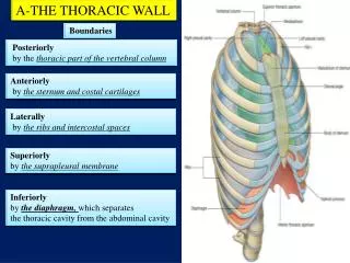
The Thoracic Wall. Surface Anatomy : Before starting your dissection, inspect and palpate the following surface landmarks with the cadaver in the supine position; later examine the same landmarks on yourself. Identification of landmarks is easier on the living than on the preserved cadaver.

E N D
Surface Anatomy: Before starting your dissection, inspect and palpate the following surface landmarks with the cadaver in the supine position; later examine the same landmarks on yourself.Identification of landmarks is easier on the living than on the preserved cadaver.
1.Suprasternal Notch.2.Sternal Angle (Angle of Louis).3. Xiphisternal Joint.4. Subcostal Angle.5. Costal Margin.6. Clavicle.7. Ribs.8. Nipple.9. Axillary Folds.
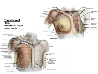
skin incisionMake a skin incision along the full length of both clavicle. Then incise the skin in the midline from the suprasternal notch to the xiphisternal junction; continue the incision along the costal margin to the midaxillary line on each side. Make circular incisions around the nipples.Reflect theskin from the midline anteriorly to the midaxillary line on each side. Leave the large skin flaps attached at the midaxillary lines.
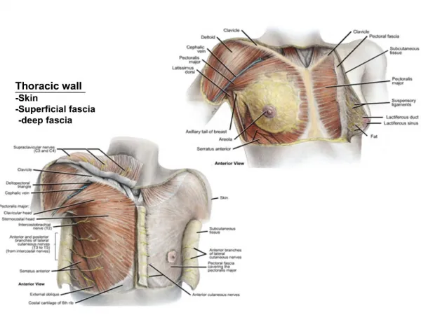
Mammary Gland. The mammary gland lies in the superficial fascia covering the anterior chest wall. In the child and in men it isrudimentary. In the femal after puberty it enlarges and assumesits hemispherical shape. In the youngadult female it overlies the second to the sixth ribs and their costal cartilages and extends from the lateral margin of the sternum to the midaxillary line. Its upper lateraledge extends round the lower border of the pectoralis major and enters the axilla.
In middle-aged multiparous women the breast may be large and pendulous. In older women past the menopause the adipose tissue of the breast may become reduced in amount and hemispherical shape lost; thebreasts then become smaller, and the overlying skin is wrinkled.
Examine the mammary gland in the male and the female. Notethat the nipple is surrounded by a pigmented area called the areola. Incise the nipple in the female and extend the incision into the breast. Observe that the breast tissues divided up into 15 to 20 compartments by fibrous septa and that the greater part of the breast substance is made up of fatty tissue. After removing thefat with the scalpel handle, identify one or two of the lactiferous ducts and trace them to the nipple. Identify the thin sheet of muscle, the platysma, asit arises from the fascia covering the pectoralis major and passes superiorly over the clavicle into the neck.
Cutaneous nerves and blood vessels. Identify the supraclavical nerves as they descend from the neck beneath the platysma. These supply the skin of the shoulder and upper part of the thorax down to the level of the second costal cartilage.
Identify the anterior and lateral cutaneous branches of an intercostal nerve.With the scalpel handle, firmly stroke the superficial fascia along the course of these nerves, thus removing the overlying fat. Very often the small blood vessels that accompany the nerves are firstidentified and lead the dissector to the nerves. Do not dissect out all the cutaneous nerves, but spend some time tracing out a representative sample.
Pectoralis Major. Expose and clean the entir anterior surface of the pectoralis major muscle. Identify the clavicular and sternocostal origins of this muscle. Follow the tendon to its insertion in the floor of the bicipital groove of the humerus. Recognize the deltopectoral triangle, an interval between the deltoid and pectoralis major muscles and the clavicle. Identify the cephalic vein as it enters the triangle from the upper limb.
Serratus Anterior.Expose the digitations of origin of the serratus anterior from the upper eight ribs. This important musclepasses round the thoracic wall to be inserted into the medial border of the scapula. Identify the anterior layer of the rectus sheath..
Cut the pectoralis major free from its origins on the clavicle, sternum, ribs, and anterior layer of the rectus sheath. Reflect the pectoralis major laterally to expose the underlying pectoralis minor, the clavipectoral fascia, and the subclavius muscle. Identify the lateral pectoral nerve as it pierces the clavipectoral fascia to enter the pectoralis major muscle. Also identify the medial pectoral nerve as it pierces the pectoralis minor to enter the pectoralis major.
Pectoralis Minor.Clean the origin of the pectoralis minor from the third, fourth, and fifth ribs. Trace the fibers laterally as they converge to be inserted into the medial border of the coracoid process.
Clavipectoral Fascia.Identify this strong sheet of connective tissue, which is split above to enclose the subclavius muscle and is attatched to the clavicle. Inferiorly, it splits to enclose the pectoralis minor muscle and continues downward to join the fascial floor of the armpit.
Identify the thoracoacromialvessels and cephalic vein as they pierce the clavipectoral fascia. Cut the pectoralis minor muscle free from its origin on the ribs and reflect it laterally. Recognize the lateral thoracic vessels along the lateral border of the pectoralis minor muscle. Do not attempt to dissect the contents of the axilla. This area forms part of the dissection of the upper limb.