THORACIC CAGE and WALL
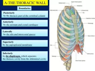
THORACIC CAGE and WALL. SURFACE LANDMARKS. Lines of Orientation. Refer: Text: pp 91-92 (91) Syllabus: p 44 Midsternal line Midclavicular line Anterior axillary line Posterior axillary line. Lines of Orientation. Midaxillary line Scapular line. Surface Landmarks. Jugular notch


THORACIC CAGE and WALL
E N D
Presentation Transcript
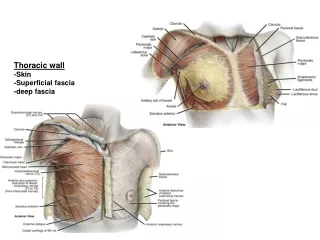
THORACIC CAGEand WALL SURFACE LANDMARKS
Lines of Orientation • Refer: Text: pp 91-92 (91) Syllabus: p 44 • Midsternal line • Midclavicular line • Anterior axillary line • Posterior axillary line
Lines of Orientation • Midaxillary line • Scapular line
Surface Landmarks • Jugular notch • Sternal angle: Marks rib 2 Lies at level of T4-5 IV disc • Apex heart beat: Left IC space 5; midclavicular line • Aortic valve: Right IC space 2
Surface Landmarks • Infrasternal angle: Important in CPR. Depression between is the infrasternal fossa. Xiphisternal joint within the fossa is at the level of the body of T9.
Surface Landmarks • Pulmonary valve Left IC space 2 • Tricuspid valve Right IC space 5 • Bicuspid valve Left IC space 5 • Bifurcation of trachea Vertebral level T4-5
Surface Landmarks • Posterior thorax: scapula: Scapular spine is at level of 3rd rib and T2 vertebra. Inferior scapular angle is at the level of the 7th rib, the spine of T7 or the body of T9. • Costal margin: Superior part is marked by the 7th cartilage. Inferior part is marked by the 10th cartilage
Cutaneous innervation • Clavicular and scapular regions: C3-4 • Region of xiphoid process: T6 • Anterior thorax: T1-T6 • Upper extremity: C5-T1 • Lower thorax and anterior abdomen: T7-T12
Structure of the Thoracic Cage • Twelve thoracic vertebrae • Twelve pairs of ribs and their costal cartilages • Sternum • Intercostal muscles
Superior Boundary (outlet) • First thoracic vertebra • First pair of ribs and costal cartilages • Manubrium • Refer to Fig. 14, p. 53 in syllabus
Inferior Boundary (outlet) • Twelfth thoracic vertebra • Twelfth pair of ribs and costal cartilages • Xiphisternal joint • Refer to Fig. 14, p. 53 in syllabus
Bones of the Thoracic Cage • Manubrium • Body of sternum: Four sternebrae • Xiphoid process • Manubriosternal joint: Symphysis Synostosis after age 30 in some
Ribs • Costa: = Rib bone + its cartilage • Vertebrosternal ribs: 1-7 • Vertebrochondral ribs: 8-10 • Vertebral ribs (floating): 11-12
Typical Rib • Head: Two articular facets for articulation with costal demifacets on adjacent thoracic vertebrae. Bony crest between facets for attachment to intervertebral disc.
Typical Rib • Neck: Narrow portion between head and tubercle. • Tubercle: Articulates with transverse process of vertebra with same number.
Typical Rib • Shaft • Sternal extremity • Angle
Typical Rib • Costal groove: Contains superior to inferior: Intercostal vein Intercostal artery Intercostal nerve • Costal cartilage
First Rib • Flattened in a horizontal (transverse) plane. • Scalene tubercle: For insertion of scalenus anterior muscle. • Shallow groove for subclavian vein: Anterior to tubercle • Shallow groove for subclavian artery: Posterior to tubercle
Thoracic Vertebra • Heart-shaped centrum (body) • Centrum notched on left side for descending aorta • Intervertebral discs = ¼ of total length of thoracic vertebral region • Superior and inferior costal demifacets: (on typical vertebrae (T2 – T9)
Thoracic Vertebra • Vertebra prominens (C7): First spinous process to be palpated. • T1 spinous process most prominent
Atypical Thoracic Vertebra • T1: Superior costal facet (not a demifacet). • T10: One pair of costal facets located partly on body and partly on pedicle. • T11 and T12: One pair of costal facets located on pedicles.
Typical Rib Articulation • Head articulates with demifacets of its own number vertebra and the vertebra above as well as the intervertebral disc between: Tubercle articulates with transverse process of same number vertebra.