Download
1 / 18
180 likes | 362 Vues
Sagittal, T1-weighted MR scan of thoracic cage. Axial CT scan of lungs at hilum. Posteroanterior (PA) chest radiograph showing a right upper lobe tumor.

E N D
Posteroanterior (PA) chest radiograph showing a right upper lobe tumor.
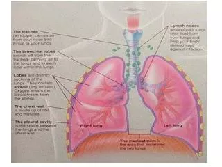
Coronal view with distribution of mediastinal lymph nodes. L, Left; R, right; 1, highest mediastinal nodes; 2, upper paratracheal nodes; 3,prevascular and retrotracheal nodes; 4, lower paratracheal (including azygos) nodes; 5,subaortic nodes; 6,paraaortic nodes; 7,subcarinal; 8,paraesophageal nodes; 9, pulmonary ligament; 10,hilar nodes; 11,interlobar nodes; 12, lobar nodes; 13, segmental nodes; 14, segmental nodes.
Digitally reconstructed radiograph for parallel-opposed fields for right lung tumor with extension across the midline.
Isodose distribution of anteroposterior (AP)/posteroanterior (PA) fields. C,Dose-volume histogram of AP/AP fields.