Download
1 / 15
170 likes | 182 Vues
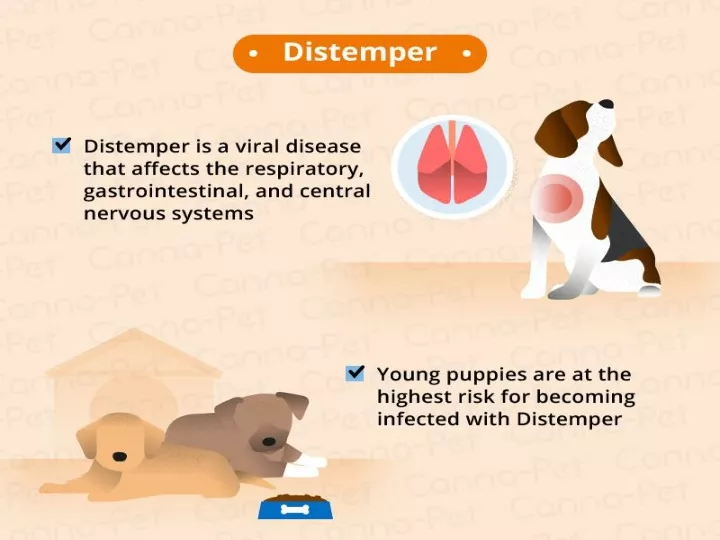
highly contagious disease caused by a virus that attacks the respiratory , gastrointestinal , and often, the nervous systems of puppies and dogs. Infected dogs shed the virus through bodily secretions and excretions , especially respiratory secretions .

E N D
highly contagious disease caused by a virus that attacks the respiratory, gastrointestinal, and often, the nervous systems of puppies and dogs.
Infected dogs shed the virus through bodily secretions and excretions, especially respiratory secretions. • The primary mode of transmission is airborne viral particles that dogs breathe in. • Dogs in recovery may continue to shed the virus for several weeks after symptoms disappear, but they no longer shed the virus once they are fully recovered. There is no cure.
History • The first case of CDV was described in 1905 by Dr. Henri Carre. • First thought to be related to the plague or Typhus (Rickettsiae) • Despite vaccine development Canine Distemper is still very prevalent today.
Etiology • It is caused by the canine distemper virus (CDV). • CDV is fairly common in wildlife. • The development of a vaccine in the early 1960s led to a dramatic reduction in the number of infected domestic dogs. • It tends to occur now only as sporadic outbreaks. • Pansystemic disease
Susceptible hosts • Puppies from 3- 6 months old are susceptible • Can infect any of the canids: • Dogs • Wolves • Foxes • Others: ferrets,
Transmission • An infected dog usually infects another by coughing infected respiratory secretions. • The virus can be shed in most all other bodily • secretions including urine. • The virus enters the body via the nose or mouth and begins to replicate. • Canine Distemper is NOT zoonotic as its name implies, will only infect other canines.
Clinical Forms of CD (5 Forms) 1-Pulmonary(Coryza like syndrome Coughing, Pneumonia) 2-Ocular (swollen eye lids, Conjunctivitis) 3-Digestive form(Vomiting, Abdominal pain, Diarrhoea) 4-Nervous form(Chewing movements, Convulsion) 5-Cutaneous form(Appearance of rash, vesicles, and pustules, the skin of foot pads and nose may become hard due to hyperkeratosis and condition called as “hard pad disease”)
Clinical Signs gggs • Begin with: • -Ocular and nasal discharge • -Fever (usually goes • unnoticed) • -Poor appetite • -Coughing • - Pneumonia • Mucosal Phase: • - Vomiting • - Diarrhea • - Callusing of the foot pads • (hard pad disease.: hyperkeratosis) • - Enamel hypoplasia (perinatal)
Neurologic Phase: • Seizures • (as convulsions, sensory disturbances, or loss of • consciousness) resulting from an abnormal • electrical discharge in the brain • Tremors (myoclonus) spasmodic jerky contraction of groups of muscles. • Imbalance • Limb weakness • Death
Diagnosis • History of immunizationagainst Canine distemper • In most cases CDV is a “clinical diagnosis” • Inclusion bodies • Inclusion bodies are actual clumps of the virus that are visible under a microscope. • Immunocytology: Test in which antibodies against distemper are tagged with fluorescent markers. Antibodies then bind to the virus if present and dye the inclusion body a glow in the dark fluorescent color.
Pathological Lesions • Pathological Lesions of CDV include: • Pulmonary congestion • Consolidation leading to focal pneumonitis. • enlarged spleen. • Bronchial Epithelium Inclusions • Lung Lesion
Treatment • The best treatment is the animals own immune response. • There are no antiviral drugs that exist to effect canine distemper, so we treat symptomatically • Antibiotics are administered for secondary bacterial infections • Airway dilators are used as needed • IV fluids are given for patients with diarrhea to prevent dehydration
Prognosis • Canine Distemper is fatal in over 50% of adult dogs who contract the virus and over 80% of puppies (90% mortality). • Death can occur between two weeks and 3 months after infection. • Main cause of death is from complications to central nervous system. • For patients in further stages of neurologic dysfunction, euthanasia is usually recommended.
Prevention • VACCINATE!!!VACCINATE!!! VACCINATE!!! • The distemper vaccine is a modified live virus and induces immune response. • Puppies should be vaccinated initially at 6-8 weeks and every 2-4 weeks after until they reach about 16 weeks of age. • Yearly Boosters! • Maintaining a sanitary environment is crucial in controlling an outbreak. • Bleach, Roccal, etc. instantly kill the virus. • Dogs with disease should be quarantined and is isolated from other patients/pets.