Download
1 / 72
780 likes | 1.85k Vues
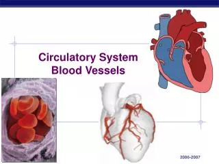
The Circulatory System Blood Vessels. Anatomy & Physiology II Chapter 15. The Vascular System. Closed system Blood vessels Four heart chambers. Blood Vessels. Delivery system of dynamic structures that begins and ends at the heart

E N D
The Circulatory SystemBlood Vessels Anatomy & Physiology II Chapter 15
The Vascular System • Closed system • Blood vessels • Four heart chambers
Blood Vessels • Delivery system of dynamic structures that begins and ends at the heart • Arteries: carry blood away from the heart; oxygenated except for pulmonary circulation and umbilical vessels of a fetus • Capillaries: contact tissue cells and directly serve cellular needs • Veins: carry blood toward the heart
Blood Vessels Five types of blood vessels • Arteries • Arterioles • Capillaries • Venules • Veins
Blood Circuits Two groups of blood vessels • The pulmonary circuit • Pulmonary artery and its branches • Capillaries in lungs • Pulmonary veins • The systemic circuit • Aorta • Systemic capillaries • Systemic veins
Blood flow in a closed system of vessels Oxygen content changes as blood flows through the capillaries. Zooming In • Judging from color coding, which vessels pick up oxygen? Which vessels release oxygen?
Vessel Structure Three tunics (coats) of arteries and veins • Inner (endothelium) • Middle (smooth [voluntary] muscle) • Controlled by autonomic nervous system • Thinner in veins • Outer (supporting connective tissue)
Sections of small blood vessels Drawings show the thick wall of an artery, the thin wall of a vein, and the single-layered wall of a capillary. A venous valve also is shown. The arrow indicates the direction of blood flow. Zooming In: Which vessels have valves that control blood flow?
Systemic Arteries The aorta • Largest artery • Receives blood from left ventricle • Branches to all organs
The Aorta and Its Parts • Ascending aorta • Aortic arch • Thoracic aorta • Abdominal aorta
The aorta and its branches Zooming In: How many brachiocephalic arteries are there?
Principal systemic arteries Zooming In: How many brachiocephalic arteries are there?
Branches of the Ascending Aorta and Aortic Arch • Ascending aorta • Left and right coronary arteries • Aortic arch • Brachiocephalic artery • Right subclavian artery • Right common carotid artery • Left common carotid artery • Left subclavian artery
Branches of the Thoracic Aorta • Branches to chest wall, esophagus, and bronchi • Intercostal arteries
Branches of the Abdominal Aorta • Celiac trunk • Left gastric artery • Splenic artery • Hepatic artery • Superior mesenteric artery • Inferior mesenteric artery • Paired lateral branches • Phrenic arteries • Suprarenal arteries • Renal arteries • Ovarian and testicular arteries • Lumbar arteries
The Iliac Arteries and Their Subdivisions • Internal iliac arteries • External iliac arteries • Femoral artery • Popliteal artery • Tibial arteries • Dorsalis pedis
Arteries That Branch to the Arm and Head • External carotid artery • Internal carotid artery • Subclavian artery • Vertebral artery • Axillary artery • Brachial artery • Radial artery • Ulnar artery
Anastomoses Communication between two vessels • Circle of Willis • Superficial palmar arch • Mesenteric arches • Arterial arches
Arteries that supply the brain The bracket at right groups the arteries that make up the circle of Willis.
Arterioles • Smallest arteries • Lead to capillary beds • Control flow into capillary beds via vasodilation and vasoconstriction
Systemic Veins • Superficial veins • Cephalic, basilic, median cubital veins • Saphenous veins • Deep veins • Femoral and iliac vessels • Brachial, axillary, subclavian vessels • Jugular veins • Brachiocephalic vein
The VenaeCavae and Their Tributaries • Superior vena cava • Head, neck, upper extremities • Azygos vein • Chest wall • Inferior vena cava • Right, left veins from paired parts, organs • Unpaired veins from spleen, digestive tract
Principal systemic veins Zooming In:How many brachiocephalic veins are there?
Venous Sinuses • Coronary sinus • Cranial venous sinuses • Cavernous sinuses • Petrosal sinuses • Superior sagittal sinus • Confluence of sinuses • Transverse sinuses (lateral sinuses)
Cranial venous sinuses The inset shows the paired transverse sinuses, which carry blood from the brain to the jugular veins.
The Hepatic Portal System Carries blood from abdominal organs to liver • Superior mesenteric vein • Splenic vein • Gastric, pancreatic, inferior mesenteric veins • Sinusoids
Hepatic portal system Veins from the abdominal organs carry blood to the hepatic portal vein leading to the liver. Arrows show the direction of blood flow. Zooming In: What vessel do the hepatic veins drain into?
Circulation Physiology • Blood exchanges oxygen, carbon dioxide, other substances generated by cells • Tissue fluid (interstitial fluid) is exchange medium
Connection between small blood vessels through capillaries The blood delivers oxygen (O2) to the tissues and picks up carbon dioxide (CO2) for transport to the lungs. Note the lymphatic capillaries, which aid in tissue drainage.
Capillaries • Microscopic blood vessels • Walls of thin tunica intima, one cell thick • Pericytes help stabilize their walls and control permeability • Size allows only a single RBC to pass at a time
Capillaries • In all tissues except for cartilage, epithelia, cornea and lens of eye • Functions: exchange of gases, nutrients, wastes, hormones, etc.
Capillaries • Three structural types • Continuous capillaries • Fenestrated capillaries • Sinusoidal capillaries (sinusoids)
Continuous Capillaries • Abundant in the skin and muscles • Tight junctions connect endothelial cells • Intercellular clefts allow the passage of fluids and small solutes • Continuous capillaries of the brain • Tight junctions are complete, forming the blood-brain barrier
Pericyte Red blood cell in lumen Intercellular cleft Endothelial cell Basement membrane Tight junction Pinocytotic vesicles Endothelial nucleus Continuous capillary: Least permeable, and most common (e.g., skin, muscle).
Fenestrated Capillaries • Some endothelial cells contain pores (fenestrations) • More permeable than continuous capillaries • Function in absorption or filtrate formation (small intestines, endocrine glands, and kidneys)
Pinocytotic vesicles Red blood cell in lumen Fenestrations (pores) Endothelial nucleus Intercellular cleft Basement membrane Endothelial cell Tight junction Fenestrated capillary: Large fenestrations (pores) increase permeability. Occurs in special locations (e.g., kidney, small intestine).
Sinusoidal Capillaries • Fewer tight junctions, larger intercellular clefts, large lumens • Usually fenestrated • Allow large molecules and blood cells to pass between the blood and surrounding tissues • Found in the liver, bone marrow, spleen
Endothelial cell Red blood cell in lumen Large intercellular cleft Tight junction Nucleus of endothelial cell Incomplete basement membrane Sinusoidal capillary: Most permeable. Occurs in special locations (e.g., liver, bone marrow, spleen).
Blood Flow Through Capillary Beds • Precapillary sphincters regulate blood flow into true capillaries • Regulated by local chemical conditions and vasomotor nerves
Vascular shunt Precapillary sphincters Thoroughfare channel Metarteriole True capillaries Terminal arteriole Postcapillary venule (a) Sphincters open—blood flows through true capillaries. Terminal arteriole Postcapillary venule (b) Sphincters closed—blood flows through metarteriole thoroughfare channel and bypasses true capillaries.
Capillary Exchange How substances move between cells and capillary blood • Diffusion • Main process • Blood pressure • Moves material into tissue fluid • Osmotic pressure • Moves material into capillaries
Factors Aiding Venous Return • Respiratory “pump”: pressure changes created during breathing move blood toward the heart by squeezing abdominal veins as thoracic veins expand • Muscular “pump”: contraction of skeletal muscles “milk” blood toward the heart and valves prevent backflow • Vasoconstriction of veins under sympathetic control
The Dynamics of Blood Flow Vasomotor center in medulla regulates vasomotor activities • Vasodilation • Vasoconstriction • Precapillary sphincter
Return of Blood to the Heart • Mechanisms that promote blood’s return to heart • Contraction of skeletal muscles • Valves • Breathing
Role of skeletal muscles and valves in blood return. Role of skeletal muscles and valves in blood return. (A) Contracting skeletal muscle compresses the vein and drives blood forward, opening the proximal valve, while the distal valve closes to prevent backflow of blood. (B) When the muscle relaxes again, the distal valve opens, and the proximal valve closes until blood moving in the vein forces it open again.
The Pulse • Ventricular contraction • Wave of increased pressure • Begins at heart and travels to arteries • Influenced by various factors • Body size • Gender • Age • Muscular activity • Emotion • Body temperature • Thyroid secretion
Blood Pressure • Force exerted by blood against vessel walls • Determined by heart’s output and resistance to blood flow
Systemic Blood Pressure • The pumping action of the heart generates blood flow • Pressure results when flow is opposed by resistance • Systemic pressure • Is highest in the aorta • Declines throughout the pathway • Is 0 mm Hg in the right atrium • The steepest drop occurs in arterioles
Arterial Blood Pressure • Systolic pressure: pressure exerted during ventricular contraction • Diastolic pressure: lowest level of arterial pressure • Pulse pressure = difference between systolic and diastolic pressure
Systemic Blood Pressure Systolic pressure Mean pressure Diastolic pressure