Download
1 / 63
630 likes | 695 Vues
Learn about the pancreas, pancreatic juice, enzymes for digesting proteins, carbohydrates, and fats, regulation of pancreatic secretion, and more.

E N D
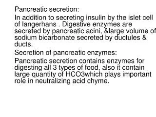
The pancreas • is a large compound gland with most of its internal structure similar to that of the salivary glands • has pancreatic digestive enzymes, secreted by pancreatic acini, and large volumes of sodium bicarbonate solution are secreted by the small ductules and larger ducts leading from the acini • the combined product of enzymes and sodium bicarbonate empties into the duodenum through the papilla of Vater, surrounded by the sphincter of Oddi. • the enzymes are secreted by gland cells at the pancreatic end of the duct system, whereas bicarbonate ions are secreted by the epithelial cells lining the ducts.
Pancreatic juice • is secreted most abundantly in response to the presence of chyme in the upper portions of the small intestine and the characteristics of the pancreatic juice are determined to some extent by the types of food in the chyme. • contains multiple enzymes for digesting all of the three major types of food: proteins, carbohydrates and fats.
The pancreatic enzymes for digesting • proteins are trypsin, chymotrypsin and carboxypolypeptidase; by far the most abundant of these is trypsin. • carbohydrates is pancreatic amylase, which hydrolyzes starches, glycogen, and most other carbohydrates (except cellulose) to form mostly disaccharides and a few trisaccharides. • fat digestion are pancreatic lipase, which is capable of hydrolyzing neutral fat; cholesterol esterase; phospholipase, which splits fatty acids from phospholipids.
When first synthesized in the pancreatic cells, the proteolytic digestive enzymes are in the inactive(trypsinogen,chymotrypsinogen and procarboxypolypeptidase) • become activated only after they are secreted into the intestinal tract. Trypsinogen is activated by an enzyme -enterokinase, which is secreted by the intestinal mucosa when chyme comes in contact with the mucosa. • Also, trypsinogen can be autocatalytically activated by trypsin that has already been formed from previously secreted trypsinogen. • Chymotrypsinogen is activated by trypsin to form chymotrypsin, andprocarboxypolypeptidase is activated in a similar manner.
It is important that the proteolyticenzymes of the pancreatic juice not become activated until after they have been secreted into the intestine because the trypsin and the other enzymes would digest the pancreas itself. • Fortunately, the same cells that secrete proteolytic enzymes into the acini of the pancreas secrete simultaneously another substance called trypsin inhibitor. This substance is formed in the cytoplasm of the glandular cells, and it prevents activation of trypsin both inside the secretory cells and in the acini and ducts of the pancreas.
The other two important components of pancreatic juice, bicarbonate ions and water, are secreted mainly by the epithelial cells of the ductules and ducts that lead from the acini. • When the pancreas is stimulated to secrete copious quantities of pancreatic juice, thebicarbonate ion concentration can rise to as high as 145 mEq/L, a value about five times that of bicarbonate ions in the plasma. • This provides a large quantity of alkali in the pancreatic juice that serves to neutralize the hydrochloric acid emptied into the duodenum from the stomach.
Regulation of pancreatic secretion Three basic stimuli are important in causing pancreatic secretion: • Acetylcholine- released from the parasympathetic vagus nerve endings and fromother cholinergic nerves in the enteric nervous system • Cholecystokinin-secreted by the duodenal and upper jejunal mucosa when foodenters the small intestine • Secretin- secreted by the duodenal and jejunal mucosa when highly acid food enters the small intestine
Regulation of pancreatic secretion • Acetylcholine and cholecystokinin, stimulate the acinar cells of the pancreas, causing production of large quantities of pancreatic digestive enzymes but relatively small quantities of water and electrolytes to go with the enzymes. • Without the water, most of the enzymes remain temporarily stored in the acini and ducts until more fluid secretion comes along to wash them into the duodenum.
Regulation of pancreatic secretion Secretin, in contrast to the first two, stimulates secretion of large quantities of water solution of sodium bicarbonate by the pancreatic ductal epithelium. When all the different stimuli of pancreatic secretion occur at once, the total secretion is far greater than the sum of the secretions caused by each one separately. Therefore, the various stimuli are said to “multiply,” or “potentiate,” one another. Thus, pancreatic secretion normally results from the combined effects of the multiple basic stimuli, not from one alone.
Regulation of pancreatic secretion Cephalic and gastric phases During the cephalic phase of pancreatic secretion, the same nervous signals from the brain that cause secretion in the stomach also cause acetylcholine release by the vagal nerve endings in the pancreas. This causes moderate amounts of enzymes to be secreted into the pancreatic acini, accounting for about 20 % of the total secretion of pancreatic enzymes after a meal
Regulation of pancreatic secretion Intestinal Phase. When chyme enters the small intestine, pancreatic s. becomes copious, mainly in response to the secretin, a polypeptide (27 amino acids), present in an inactive form, prosecretin, in so-called S cells in the mucosa of the duodenum and jejunum. When acidchyme with pH less than 4.5 to 5.0 enters the duodenum from the stomach, it causes duodenal mucosal release and activation of secretin, which is then absorbed into the blood. Secretin in turn causes the pancreas to secrete large quantities of fluid containing a high concentration of bicarbonate ion (up to 145 mEq/L) but a low concentration of chloride ion.
Regulation of pancreatic secretion • The presenceof food in the upper small intestine also causes asecond hormone, cholecystokinin, (33 AA), to be released from anothergroup of cells, the I cells, in the mucosa of the duodenum and upper jejunum. • This release of cholecystokinin results especially from the presence of proteoses and peptones (products of partial protein digestion) and long-chain fatty acidsin the chyme coming from the stomach. • Cholecystokinin, like secretin, passes by way of the blood to the pancreas but instead of causing sodium bicarbonate secretion causes mainly secretion of still much more pancreatic digestive enzymes by the acinar cells.
Bile secretion • Bile is secreted by liver cells into a number of small ducts, the bile canaliculi, which converge to form the common hepatic duct. • Bile contains six major ingredients: (1) bile salts; (2) lecithin ; (3) bicarbonate ions and other salts; (4)cholesterol; (5) bile pigments and small amounts of other metabolic end products, and (6) trace metals.
Bile secretion • Bile salts and lecithin are synthesized in the liver and help solubilize fat in the small intestine. • Bicarbonate ions neutralize acid in the duodenum, and • The last three ingredients represent substances extracted from the blood by the liver and excreted via the bile.
Bile secretion • Bile secretion, followed by excretion of cholesterol in the feces, is one of the mechanisms by which cholesterol homeostasis in the blood is maintained . • Cholesterol is insoluble in water and its solubility in bile is achieved by its incorporation into micelles (whereas in the blood, cholesterol is incorporated into lipoproteins).
Bile secretion • Bile pigments are substances formed from the heme portion of hemoglobin when old or damaged erythrocytes are digested in the spleen and liver. • The predominant bile pigment is bilirubin, which is extracted from the blood by liver cells and actively secreted into the bile. It is bilirubin that gives bile its yellow color. • After entering the intestinal tract, bilirubin is modified by bacterial enzymes to form the brownpigments that give feces their characteristic color. During their passage through the intestinal tract, some of the bile pigments are absorbed into the blood and are eventually excreted in the urine, giving urine its yellow color.
Bile secretion Like pancreatic secretions, the components of bile are secreted by two different cell types. • The bile salts, cholesterol, lecithin, and bile pigments are secreted by hepatocytes, • whereas most of the bicarbonate-rich salt solution is secreted by the epithelial cells lining the bile ducts. Secretion of the salt solution by the bile ducts, just like that secreted by the pancreas, is stimulated by secretin in response to the presence of acid in the duodenum
Bile secretion • Unlike the pancreas, whose secretions are controlled by intestinal hormones, bile salt secretion is controlled by the concentration of bile salts in the blood—the greater the plasma concentration of bile salts, the greater their secretion into the bile canaliculi. • Absorption of bile salts from the intestine during the digestion of a meal leads to their increased plasma concentration and thus to an increased rate of bile salt secretion by the liver
Bile secretion • One of the many functions of the liver is to secrete bile, normally between 600 and 1000 ml/day. • The most abundant substances secreted in the bile are bile salts, which account for about one half of the total solutes also in the bile. • In the concentrating process in the gallbladder, water and large portions of the electrolytes (except calcium ions) are reabsorbed by the gallbladder mucosa; essentially all other constituents, especially the bile salts and the lipid substances cholesterol and lecithin, are not reabsorbed and, therefore, become highly concentrated in the gallbladder bile.
Bile secretion Bile is secreted in two stages by the liver: • The initial portion is secreted by the hepatocytes; this initial secretion contains large amounts of bile acids, cholesterol, and other organic constituents. • Next, the bile flows in the canaliculi toward the interlobular septa, where the canaliculi empty into terminal bile ducts and then into progressively larger ducts, finally reaching the hepatic duct and common bile duct. From these the bile either empties directly into the duodenum or is diverted for minutes up to several hours through the cystic duct into the gallbladder
Bile secretion • In its course through the bile ducts, a second portion of liver secretion is added to the initial bile.This additional secretion is a watery solution of sodium and bicarbonate ions secreted by secretory epithelial cells that line the ductules and ducts. • This second secretion sometimes increases the total quantity of bile by as much as an additional 100 %. The second secretion is stimulated especially by secretin, which causes release of additional quantities of bicarbonate ions to supplement the bicarbonate ions in pancreatic secretion (for neutralizing acid that empties into the duodenum from the stomach
Bile secretion • When food begins to be digested in the upper gastrointestinal tract, the gallbladder begins to empty, especially when fatty foods reach the duodenum about 30 minutes after a meal. • The mechanism of gallbladder emptying is rhythmical contractions of the wall of the gallbladder, but effective emptying also requires simultaneous relaxation of the sphincter of Oddi, which guards the exit of the common bile duct into the duodenum. By far the most potent stimulus for causing the gallbladder contractions is the hormone cholecystokinin.
The gallbladder empties its store of concentrated bile into the duodenum mainly in response to the cholecystokinin stimulus that itself is initiated mainly by fatty foods. • When fat is not in the food, the gallbladder empties poorly, but when significant quantities of fat are present, the gallbladder normally empties completely in about 1 hour
The bile salts have two important actions: • a detergent action on the fat particles in the food (decreases the surface tension of the particles and allows agitation in the intestinal tract to break the fat globules into minute sizes) • even more important, bile salts help in the absorption of fatty acids, monoglycerides, cholesterol, and other lipids from the intestinal tract. ----by forming very small physical complexes with these lipids, called micelles, and they are semi-soluble in the chyme because of the electrical charges of the bile salts. The intestinal lipids are “ferried” in this form to the intestinal mucosa, where they are then absorbed into the blood.
About 94 % of the bile salts are reabsorbed into the blood from the small intestine, about one half of this by diffusion through the mucosa in the early portions of the small intestine and the remainder by an active transport process through the intestinal mucosa in the distal ileum. • They then enter the portal blood and pass back to the liver. On reaching the liver, on first passage through the venous sinusoids these salts are absorbed almost entirely back into the hepatic cells and then are resecreted into the bile.
This recirculation of the bile salts is called the enterohepatic circulation of bile salts. • The quantity of bile secreted by the liver each day is highly dependent on the availability of bile salts—the greater the quantity of bile salts in the enterohepatic circulation (usually a total of only about 2.5 grams), the greater the rate of bile secretion. • The daily rate of liver bile salt secretion is actively controlled by the availability (or lack of availability) of bile salts in the enterohepatic circulation.
Physiology of the Liver • The liver is the largest organ in the body, contributing about 2 % of the total body weight, or about 1.5 kg in the average adult human. • The basic functional unit of the liver is the liver lobule, which is a cylindrical structure several millimeters in length and 0.8 to 2 millimeters in diameter. The human liver contains 50,000 to 100,000 individual lobules.
Physiology of the Liver • The liver lobule is constructed around a central vein that empties into the hepatic veins and then into the vena cava. • The lobule is composed principally of many liver cellular plates that radiate from the central vein like spokes in a wheel. Each hepatic plate is usually two cells thick and between the adjacent cells lie small bile canaliculi that empty into bile ducts in the fibrous septa separating the adjacent liver lobules. • In the septa are small portal venules that receive their blood mainly from the venous outflow of the gastrointestinal tract by way of the portal vein. From these venules blood flows into flat, branching hepatic sinusoids that lie between the hepatic plates and then into the central vein.
Physiology of the Liver • Hepatic arterioles are also present in the interlobular septa. • These arterioles supply arterial blood to the septal tissues between the adjacent lobules and many of the small arterioles also empty directly into the hepatic sinusoids, most frequently emptying into those located about one third the distance from the interlobular septa
Physiology of the Liver • In addition to the hepatic cells, the venous sinusoids are lined by two other types of cell: • typical endothelial cells and • large Kupffer cells (or reticuloendothelial cells), which are resident macrophages that line the sinusoids and are capable of phagocytizing bacteria and other foreign matter in the hepatic sinus blood. The endothelial lining of the sinusoids has extremely large pores, some of which are almost 1 micrometer in diameter.
Physiology of the Liver • Blood flows through the liver from the portal vein and hepatic artery. The liver has high blood flow and low vascular resistance. • The pressure in the portal vein leading into the liver averages about 9 mm Hg, and the pressure in the hepatic vein leading from the liver into the vena cava normally averages almost exactly 0 mm Hg. This small pressure difference, only 9 mm Hg, shows that the resistance to blood flow through the hepatic sinusoids is normally very low.
1.The liver functions as a blood reservoir • Because the liver is an expandable organ, large quantities of blood can be stored in its blood vessels. • Its normal blood volume, including both that in the hepatic veins and that in the hepatic sinuses, is about 450 milliliters, or almost 10 % of the body’s total blood volume. • When high pressure in the right atrium causes backpressure in the liver, the liver expands, and 0.5 to 1 liter of extra blood is occasionally stored in the hepatic veins and sinuses.
2.The liver has very high lymph flow • Because the pores in the hepatic sinusoids are very permeable and allow ready passage of both fluid and proteins into the spaces of Disse, the lymph draining from the liver usually has a protein concentration of about 6 g/dl, which is only slightly less than the protein concentration of plasma; • the extreme permeability of the liver sinusoid epithelium allows large quantities of lymph to form. Therefore, about half of all the lymph formed in the body under resting conditions arises in the liver.
3.Regulation of liver mass— regeneration • remarkable ability to restore itself after significant hepatic tissue loss from either partial hepatectomy or acute liver injury, as long as the injury is uncomplicated by viral infection or inflammation. • Partial hepatectomy, in which up to 70% of the liver is removed, causes the remaining lobes to enlarge and restore the liver to its original size. This regeneration is remarkably rapid and requires only 5 to 7 days in rats. • During liver regeneration, hepatocytes are estimated to replicate once or twice, and after the original size and volume of the liver are achieved, the hepatocytes revert to their usual state.
4.Hepatic macrophage system serves a blood-cleansing function • Blood flowing through the intestinal capillaries picks up many bacteria from the intestines. • Special high-speed motion pictures of the action of Kupffer cells, the large phagocytic macrophages that line the hepatic venous sinuses, have demonstrated that these cells efficiently cleanse blood as it passes through the sinuses; • when a bacterium comes into momentary contact with a Kupffer cell, in less than 0.01 second the bacterium passes inward through the wall of the Kupffer cell to become permanently lodged therein until it isdigested.
5.Metabolic functions of the liver The most of water-soluble nutrients and water-soluble vitamins and minerals absorbed from the small intestine are transported via the portal blood to the liver. The nutrients transported in portal blood include amino acids, monosaccharides and fatty acids (predominantly short- andmedium-chain forms). Short-chain fatty acids are largely derived from the fermentation of dietary fibers by bacteria in the colon.
5.Metabolic functions of the liver a)Carbohydrate metabolism- the liver performs the following functions: 1. Storage of large amounts of glycogen 2. Conversion of galactose and fructose to glucose 3. Gluconeogenesis 4. Formation of many chemical compoundsfrom intermediate products of carbohydrate metabolism
5.Metabolic functions of the liver • The liver is especially important for maintaining a normal blood glucose concentration. • Storage of glycogen allows the liver to remove excess glucose from the blood, store it, and then return it to the blood when the blood glucose concentration begins to fall too low. • Thisis called the glucose buffer functionof the liver
5.Metabolic functions of the liver b)Fat metabolism Although most cells of the body metabolize fat, certain aspects of fat metabolism occur mainly in the liver. Specific functions are: 1. Oxidation of fatty acids to supply energy for other body functions 2. Synthesis of large quantities of cholesterol,phospholipids, and most lipoproteins 3. Synthesis of fat from proteins and carbohydrates
5.Metabolic functions of the liver • To derive energy from neutral fats, the fat is first split into glycerol and fatty acids; then the fatty acids are split by beta-oxidation into two-carbon acetyl radicals that form acetyl coenzyme A (acetyl-CoA). • This can enter the citric acid cycle and be oxidized to liberate tremendous amounts of energy. • Beta-oxidation can take place in all cells of the body, but it occurs especially rapidly in the hepatic cells
5.Metabolic functions of the liver • About 80 % of the cholesterol synthesized in the liver is converted into bile salts, which are secreted into the bile; the remainder is transported in the lipoproteins and carried by the blood to the tissue cells everywhere in the body. • Phospholipids are likewise synthesizedin the liver and transported principally in the lipoproteins. • Both cholesterol and phospholipids are used by the cells to form membranes, intracellular structures, and multiple chemical substances that are important to cellular function
5.Metabolic functions of the liver c)Protein metabolism The body cannot dispense with the liver’s contribution to protein metabolism for more than a few days without death ensuing. 1. Deamination of amino acids 2. Formation of urea for removal of ammonia from the body fluids 3. Formation of plasma proteins 4. Interconversions of the various amino acids and synthesis of other compounds from amino acids