Download

1 / 19
650 likes | 2.24k Vues
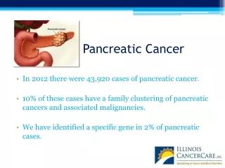
Pancreatic cancer. By Linda Sircy. Where is the pancreas?. Between the stomach and spine Lies partially behind the stomach and rests in the curve of the small intestine. http:// www.webmd.com /digestive-disorders/picture-of-the-pancreas. Statistics. The ACS estimates:

E N D
Pancreatic cancer By Linda Sircy
Where is the pancreas? • Between the stomach and spine • Lies partially behind the stomach and rests in the curve of the small intestine http://www.webmd.com/digestive-disorders/picture-of-the-pancreas
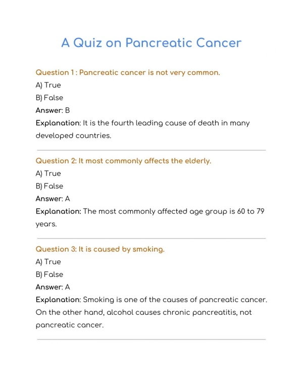
Statistics The ACS estimates: • About 45,220 people (22,740 men and 22,480 women) will be diagnosed with pancreatic cancer • About 38,460 people (19,480 men and 18,980 women) will die of pancreatic cancer • Rates of pancreatic cancer have been slowly increasing over the past 10 years • The lifetime risk of developing pancreatic cancer is about 1 in 78.
Exocrine tumors • Most common type • Benign cysts and benign tumors (cystadenomas) can occur, but most are malignant. • About 95% are adenocarcinomas • Less common types include: • Adenosquamous carcinomas • Squamous cell carcinomas • Signet ring cell carcinomas • Undifferentiated carcinomas • Undifferentiated carcinomas with giant cells • Solid pseudopapillary neoplasms of the pancreas • Ampullarycancer (or carcinoma of the ampulla of Vater)
Endocrine tumors • Known as pancreatic neuroendocrine tumors (NETs), or islet cell tumors • Subtypes include: • Insulinomas** • Gastrinomas** • Glucagonomas • Somatostatinomas • VIPomas(Vasoactive Intestinal Peptide) • Ppomas (Pancreatic Polypeptide) • Carcinoid tumors
Risk factors • Age • Gender • Race • Tobacco use • Obesity • Diabetes • Chronic pancreatitis • Cirrhosis of the liver • Occupational exposure • Stomach problems • Diet • Coffee • Alcohol
Risk factors Family history/Genetic syndromes • Inherited gene mutations may cause as many as 10% of pancreatic cancers: • Hereditary breast and ovarian cancer syndrome • Familial melanoma • Familial pancreatitis • Hereditary non-polyposis colorectal cancer (HNPCC), also known as Lynch syndrome. • Peutz-Jeghers syndrome (PJS), also linked with polyps in the digestive tract and several other cancers • Von Hippel-Lindausyndrome • Pancreatic neuroendocrine tumors and cancers can also be caused by a genetic syndrome, such as: Neurofibromatosis type 1 and Multiple endocrine neoplasiatype 1
Signs and symptoms • Exocrine tumors • Jaundice • Darkening urine • Abdominal or back pain • Weight loss and poor appetite • Digestive problems • Gallbladder enlargement • Blood cots or fatty tissue abnormalities • Diabetes • Endocrine tumors • Stomach ulcers, abdominal pain, nausea • Decreased appetite, weight loss, malnutrition, digestion problems • Diabetes • Diarrhea, gallbladder issues, jaundice, dark urine • Fainting, coma, seizures • Rapid heart rate, weakness, shortness of breath, confusion, sweating
Signs and symptoms • Because of the pancreas' deep location, tumors are rarely palpable through the abdomen. • Many symptoms of pancreatic cancer often do not appear until the tumor grows large enough to interfere with the function of nearby structures: • Stomach • Duodenum • Liver • Gallbladder
Diagnostic tests • CT scan • CT-guided needle biopsy • MRI • Somatostatin receptor scintigraphy • Used for diagnosing NETs • Positron emission tomography (PET) scan • Used to look at spread from exocrine tumors • Ultrasonography • Endoscopic ultrasound • Laparoscopy • X-ray • Endoscopic retrograde cholangiopancreatography (ERCP) • Percutaneous transhepatic cholangiography (PTC) • Angiography • Blood tests • Used for diagnosing NETs • Biopsy
Grading • Pancreatic cancer does not use a specific grading system, so it follows the general system: • GX: Undetermined grade • G1: Well differentiated or low grade • G2: Moderately differentiated or intermediate grade • G3: Poorly differentiated or high grade • G4: Undifferentiated or high grade
Staging • Stage 0 • Stage I • IA • IB • Stage II • IIA • IIB • Stage III • Stage IV
Other terms: • Another factor in staging pancreatic cancers is the extent of resection: • From R0, where all visible and microscopic tumor was removed… • To R2, where some visible tumor could not be removed • Some doctors use a simpler staging system, dividing cancers into groups based on likelihood of surgical removal: • Resectable • Locally advanced (or unresectable) • Metastatic
Treatment options • Surgery • Palliative surgery • Radiation • Chemotherapy • Biologic therapy • Ablative techniques
References • http://pathology.jhu.edu/pc/BasicOverview1.php • http://www.cancer.org/cancer/pancreaticcancer/index • http://www.cancer.gov/cancertopics/types/pancreatic • http://www.mayoclinic.com/health/pancreatic-cancer/DS00357 • http://www.cancer.gov/cancertopics/factsheet/detection/tumor-grade • http://www.cancer.gov/cancertopics/pdq/treatment/pancreatic/Patient/page2 • http://www.upmccancercenter.com/pdq_xml/cancer.cfm?id=105