Download
1 / 155
1.62k likes | 2.68k Vues
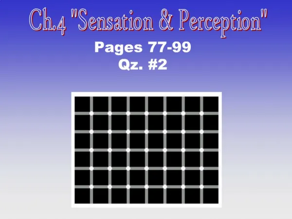
Chapter 4 Sensation and Perception. Sensation and Perception. Sensation is the conversion of energy from the environment into a pattern of response by the nervous system. Perception is the interpretation of that information. Module 4.1. Vision. Sensing the World Around Us.

E N D
Sensation and Perception • Sensation is the conversion of energy from the environment into a pattern of response by the nervous system. • Perception is the interpretation of that information.
Module 4.1 • Vision
Sensing the World Around Us • Stimuli are energies in the environment that affect what we do. • Receptors are the specialized cells in our bodies that convert environmental energies into signals for the nervous system.
The Detection of Light • Light is the stimulus that the visual system is designed to detect. • Visible light is just one very small portion of the electromagnetic spectrum, which is the continuum of all the frequencies of radiated energy. • The human eye is designed to detect energy in the wavelengths from 400 to 700 nm.
Figure 4.2 The lens gets its name from Latin for lentil, referring to its shape—an appropriate choice, as this cross section of the eye shows. The names of other parts of the eye also refer to their appearance.
The Structure of the Eye • The pupil is an adjustable opening in the eye through which light enters. • The iris is the structure on the surface of the eye, surrounding the pupil, and containing the muscles that make the pupil dilate or constrict. • The iris gives your eye its characteristic color, too.
The Structure of the Eye • The cornea is a rigid, transparent structure on the very outer surface of the eyeball. It focuses light by directing it through the pupil. • When the light goes through the pupil, it is directed to the lens. • The lens is a flexible structure that can vary in thickness, enabling the eye to accommodate, adjusting its focus for objects at different distances.
The Structure of the Eye • The lens directs the light through a clear, jellylike substance called the vitreous humor to the back of the eyeball. • At the back of the eye is the retina, the structure containing the visual receptors.
Common Disorders of Vision • Presbyopia develops as humans age because the lens decreases in flexibility, resulting in a reduced ability to focus on nearby objects. • Elongated eyeballs cause myopia, so that the person can focus well on nearby objects, but not distant ones. This condition is also called nearsightedness. • Flattened eyeballs cause hyperopia, so that the person can focus well on distant objects, but not on nearby ones. This is also called farsightedness.
Figure 4.3 The flexible, transparent lens changes shape so that objects (a) far and (b) near can come into focus. The lens bends entering light rays so that they fall on the retina. In old age the lens becomes rigid, and people find it harder to focus on nearby objects
Common Disorders of Vision • Glaucoma is a condition caused by increased pressure within the eyeball, causing damage to the optic nerve and loss of peripheral vision. • A cataract is a disorder in which the lens of the eye becomes cloudy. This disorder is treated by removing and replacing the actual lens with a contact lens.
Concept Check What happens if a person with normal vision puts on contact lenses designed for a person with myopia? His vision, especially his near vision, will be blurry.
The Visual Receptors • The retina contains two types of specialized neurons, the rods and the cones. • Rods far outnumber cones in the human eye. • About 5-10% of the visual receptors in the human retina are cones.
The Visual Receptors • The cones are utilized in color vision, daytime vision and detail vision. • The rods are adapted for vision in dim light. • Species that are active at night have few cones and many rods, giving them particularly good night vision.
The Visual Receptors • The fovea is the center of the human retina, and the location of the highest proportion of cones. • It is the area of the eye with the greatest acuity. • Rods are more plentiful in the periphery of the retina.
Concept Check If you see a brightly colored object in the periphery of your vision, the colors will not seem very bright at all. Why is this? You have mostly rods in the periphery of your retina, thus a more limited ability to detect color.
Dark Adaptation • Most humans require one or two minutes to see in the dark. This process of gradual improvement is called dark adaptation. • Exposure to light causes molecules of retinaldehydes to be chemically altered and stimulate the visual receptors.
Dark Adaptation • In conditions of normal daytime light, these molecules are depleted and regenerated at about the same rate, so the amount available in the retina is balanced and level of visual sensitivity is constant. • In dim light or darkness the receptors regenerate these molecules without any subsequent depletion, and so the increase in available retinaldehydes also enhances dark adaptation.
Dark Adaptation • Cones and rods perform this regeneration at different rates. • Although cones regenerate their retinaldehydes more quickly, they are also being more heavily used in the daytime. • By the time you need enhanced night vision, the rods have fully regenerated their supply of retinaldehydes. • The relative abundance of rods, and their undisturbed regeneration of the chemical, gives them a much higher level of sensitivity to faint light
Figure 4.8 These graphs show dark adaptation to (a) a light you stare at directly, using only cones, and (b) a light in your peripheral vision, which you see with both cones and rods. (Based on E. B. Goldstein, 1989)
Concept Check: It is said that dogs and cats can see in the dark – do you think this is really true? Although these animals have much better vision in dim light than we do, there must be some light present for the rods to function.
Concept Check In the daytime which predominates, the fovea or periphery of the eye? Unless you walk into a dark room, you will be using the fovea, because cones are the receptors for daytime (well-lighted) vision.
The Visual Pathway • The visual receptors send their impulses away from the brain, toward the center of the eye. • First the bipolar cells gather the impulses from the rods and cones. • Then the bipolar cells make synaptic contacts with ganglion cells.
Figure 4.7 Because so many rods converge their input into the next layer of the visual system, known as bipolar cells, even a small amount of light falling on the rods can stimulate the bipolar cells. Thus, the periphery of the retina, with many rods, has good perception of faint light. However, because bipolars in the periphery get input from so many receptors, they have only imprecise information about the location and shape of objects.
The Visual Pathway • The axons of the ganglion cells join together to form the optic nerve, which makes a “U-turn” and exits the eye. • There are no photoreceptors at the point at which the nerve leaves the eye. This is called the blind spot. • You are not aware of your blind spot because information from the retina of each eye “fills in” the blind spot in the other eye. This integration occurs in the visual cortex.
The Visual Pathway • At the optic chiasm, half of each optic nerve crosses to go to the opposite side of the brain. • At this point the axons begin to separate, sending information to a number of locations in the brain. • The greatest number of axons goes to the occipital lobe via the thalamus.
Figure 4.9 Axons from cells in the retina depart the eye at the blind spot and form the optic nerve. In humans about half the axons in the optic nerve cross to the opposite side of the brain at the optic chiasm. Some optic nerve axons carry information to the midbrain; others carry it to the thalamus, which relays information to the cerebral cortex.
The Visual Pathway • The information from each retina is integrated in the visual cortex. • Each cell in the cortex receives input from both the left and the right retinas. • When the retinas are focused on the same point in space, the input from each side is easily integrated because the message is from each is almost the same.
The Visual Pathway • If the images conflict with each other, cortical cells will be alternately stimulated and inhibited as they try to integrate the information. • The alternation between seeing the conflicting information from each retina is called binocular rivalry.
Figure 4.11 To produce binocular rivalry, move your eyes toward the page until the two circles seem to merge. You will alternate between seeing red lines and green lines.
The Visual Pathway • The brain activity of the visual cortex is crucial for the sense of vision. • People with intact eyes but a damaged visual cortex lose the ability to imagine visual imagery.
Color Vision • Different wavelengths of electromagnetic energy correspond to different colors of light. • There are three kinds of cones that respond to different wavelengths. • Cells in the visual path process the information from these cones in terms of opposites.
Color Vision • The three types of information are: • Red vs. green • Yellow vs. blue • White vs. black • The cells in the cerebral cortex integrate the input from the parts of the visual field to create a color experience for the objects that we see.
Color Vision • The Young-Helmholtz theory • This is also known as the trichromatic theory. • It proposes that our receptors respond to three primary colors. • “Color vision depends on the relative rate of response by the three types of cones.”
Color Vision • Each type of cone is most sensitive to a specific range of electromagnetic wavelengths. • Short wavelengths are seen as blue. • Medium wavelengths are seen as green. • Long wavelengths are seen as red.
Figure 4.13 Sensitivity of three types of cones to different wavelengths of light. (Based on data of Bowmaker & Dartnall, 1980)
Color Vision • Each wavelength induces different levels of activity in each type of cone. • For example, light that stimulates the medium and long wavelength cones about equally will be perceived as yellow. • Light that excites all three types equally is perceived as white.
Color Vision • The Opponent-Process Theory • Trichromatic theory does not account for some of the more complicated aspects of color perception. • People experience four colors as primary – red, green, blue and yellow. • People also report seeing colored after-images after staring at an object of one color. If you stare at a red object, you tend to see a green after-image when you stop staring.
Color Vision • The Opponent-Process Theory • Because of these facts, Ewald Hering proposed that we perceive color not in terms of separate categories but rather in a system of paired opposites. • Red vs. green • Yellow vs. blue • White vs. black
Color Vision • The Opponent-Process Theory • The negative after-images that we experience after staring at objects are results of the alternating stimulation and inhibition of neurons in the visual system. • A bipolar neuron that responds strongly to yellow will be inhibited by blue. • After you’ve stared at a yellow object, your fatigued bipolar cell will behave as if it’s been inhibited, and yield a sensation of blue.
Concept Check A bipolar cell is stimulated by red wavelengths. You stare at a red object. What will happen when you stop staring? You will see a negative afterimage in green.
Concept Check Why do negative after-images that you see seem to “move around?” Because the image is in your eye, not from any object at which you are gazing.
Color Vision • The Retinex Theory • The trichromatic and opponent-process theory don’t account for our experience of color constancy. • Color constancy is the tendency of an object to appear nearly the same color even though we see it in a variety of lighting conditions.
Color Vision • The Retinex Theory • Edwin Land proposed that we perceive color because the cerebral cortex compares various retinal patterns (thus the name retina + cortex = “retinex.”) • By comparing different patterns of light from different areas of the retina, cortical cells synthesize a color perception for each area.
Color Vision • The Retinex Theory • The fact that certain types of brain damage disrupt color constancy, causing for example an object to look orange under one level or type of lighting, and red, green, yellow or even white under other conditions, is considered to be strong evidence for the Retinex theory.
Color Vision • Colorblindness • Total inability to distinguish colors is very rare except as a result of brain damage. • About 4% of all people are partly colorblind.
Color Vision • Colorblindness • Colorblindness can result from the absence of one of the three types of cones. • Colorblindness can also result when one of the three types of cones is less responsive than the other two. The color that stimulates that type of cone is seen as almost gray.
Color Vision • Colorblindness • Red-green colorblindness is the most common type. • There are two forms – protanopia, in which the afflicted person lacks long-wavelength cones, and deuteranopia, in which the person lacks medium-wavelength cones. • Yellow-blue colorblindness (known as trianopia) is very rare.