Download
1 / 110
1.13k likes | 1.72k Vues
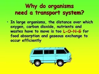
When do Organisms need Transport Systems?. We need TRANSPORT when 2 cells are far from each other materials needed to be moved from one place to another huge sum of substances to be moved. Why do Organisms need Transport Systems?.

E N D
When do Organisms need Transport Systems? • We need TRANSPORT when • 2 cells are far from each other • materials needed to be moved from one place to another • huge sum of substances to be moved
Why do Organisms need Transport Systems? • ensure a continual supply of nutrients, oxygen and other useful materials for metabolism and removal of toxic waste products produced by metabolism
small animal can undergo this by diffusion but large animal cannot, they need a transport system
blood = plasma + blood cells (straw-coloured fluid) (corpuscles) Blood Components • blood cells include red blood cell (erythrocytes), white blood cells (leucocytes ) and blood platelets where red blood cells are the most numerous blood cells and give blood the red colour
A Centrifuge for separation Plasma Blood cells Separation of blood Blood components can be separated by centrifuge
White Blood Cell Red Blood Cell 90 % Water 10% Dissolved Substances Platelets Lymphocytes Composition of Mammalian Blood Whole Blood Plasma (55% by volume) Blood Cell (45% by volume) Phagocytes
WATER (as a solvent) Blood Plasma Plasma is approximately 55% by volume and contains • SOLUBLE SUBSTANCES • hormones • protein • mineral salts • gases • dissolved food substances • metabolic wastes
Platelets White Blood Cell Red Blood Cell Blood Cells
Red Blood Cells (red corpuscles, erythrocytes) • form within bone marrow • short life span with about 120 days • old red blood cells are destroyed in liver & spleen
they have no nuclei when mature - it increases the space to carry haemoglobin • they have biconcave disc shape - which provides large surface area to diffuse oxygen
haemoglobin is an iron-containing compound and its presence is responsible for the colour of red blood cell • possess of haemoglobin • enable red blood cell to carry oxygen from lungs to all parts of the body
In lung Oxygen + haemoglobin oxyhaemoglobin In tissue Transport of Oxygen • haemoglobin has a high affinity for oxygen when the concentration of oxygen is high • change of haemoglobin to oxyhaemoglobin is accompanied by the colour change from purplish red to bright red
CO2 (in bloodstream) enzyme H+ + HCO3- hydrogen- carbonate ion H2CO3 carbonic acid CO2 + H2O (In red blood cell) HCO3- (in plasma) Transport of Carbon Dioxide CO2 (from tissue)
Investigation 12.2 To Test a Sample of Blood Plasma (chicken/pig/ox) for glucose
Fehling’s solutions A and B supernatant centrifuge boiling water chicken blood Name the supernatant obtained after centrifugation. Ans:It is plasma.
Fehling’s solutions A and B supernatant centrifuge boiling water chicken blood What does the precipitate in the centrifuge tube consist of ? Ans:The precipitate contains blood cells.
Fehling’s solutions A and B supernatant centrifuge boiling water chicken blood What happens to the supernatant when it is heated with Fehling’s solutions A and B ? Ans:The supernatant forms an orange precipitate.
White blood cells Red blood cells White Blood Cells (white corpuscles, leucocytes) • larger than red blood cells and irregular in shape • prominent nucleus • no haemoglobin • kill germs, defend against disease • two main kinds of white blood cells: phagocytes and lymphocytes
White Blood Cells - Phagocytes • made in bone marrow but different from the place where red blood cells are made • irregularly shaped nucleus • move like Amoeba • can squeeze out through the walls of capillaries into the surrounding tissues • engulf dead cells or pathogens
White Blood Cells - Lymphocytes • made in bone marrow, then migrate to lymph nodes • large nucleus which nearly fills up the cells
produce antibodies to attack germs by reaction with their surfaces and often cause them to clump together • produce antitoxins to neutralize the toxins secreted by germs
Platelets (thrombocytes) • platelets are not cells • fragments budded off from specialized cells in bone marrow • smaller than other blood cells • life-span is about 5 to 9 days • agent for initializing blood clotting
results: fibrinogen fibrin Blood Clotting • when platelets are damaged in an injury, it releases a chemical substance which starts a chain of reactions • fibrin acts like a net, trapping blood cells and plugging the wound so bleeding stops
The clotting of blood (plasma protein) • serum are yellowish fluid which is plasma without fibrinogen • when new skin formed under the scab, it loosens and comes off • clot dries up and harden to form a scab
Functions of Blood • It acts as a transport medium for oxygen, carbon dioxide, food, urea, hormones, antibodies and heat • It contains white blood cells and platelets for body defense against infection • It helps in maintaining body temperature constant
In emergencies an injured person may die... Blood Transfusion How can we save his life? Any criteria for Blood Transfusion? Donor’s blood and recipient’s blood must be compatible, otherwise, agglutination will occur which will block the blood vessels
Blood Grouping • a person’s blood group determined by the protein present on the surface of red blood cells called antigens • there are two different antigens called antigen A and antigen B. For a person in group A contains antigen A
in human, there are mainly four different blood groups called A, B, AB and O • in plasma, there may contain antibodies known as anti-A and anti-B. They will react with certain red blood cells which contain the wrong antigen
Type A Type B Type AB Type O Antigen B Antigens A and B Neither antigen A or B Antigen A Red blood cell Neither antibody A and B Antibodies A and B Antibody B Antibody A Plasma The ABO Blood Group
Blood Antigen Antibodies Recipient Group (RBC) (Plasma) A A Anti-B A & AB B B Anti-A B & AB AB A & B NO AB only O NO Both anti-A ALL & anti-B HumanBloodGroups
A Type A blood of donor Type B antibody in type A blood of recipient No agglutination B Type A blood of donor Type A antibody in type B blood of recipient Agglutination Agglutination Reaction
Donor Recipient A B Agglutination Human Blood Groups AB=universal recipient O= universal donor
Blood Vessels • there are three main kinds of vessels: arteries, veins and capillaries • arteries carry blood away from the heart while veins carry blood towards the heart
Heart Artery Vein Venule Arteriole Capillary Blood Circulation
Artery Vein Blood Vessels
Arteries carry blood away from the heart and so the blood is under high pressure wall of arteries are thick and supported with muscles and elastic fibres Artery Artery
Vein Vein • Blood pressure is much lower in vein as blood has flowed slowly through the capillaries before entering the vein • vein has larger lumen and thinner walls thanartery
Valve closed blood can’t flow back Valveopen blood can flow valves present to prevent backflow of blood and ensure that it flows towards the heart
Blood squeezed towards heart Muscle contracted Valves closed Prevent back-flow return of blood to heart is aided by contraction of body muscles as they squeeze the blood along the vein
Arteries Veins carry blood away from the heart return blood to the heart Direction of blood flow thick wall made up of muscles and elastic fibres thin wall made up of muscles and elastic fibres Wall Differences between Arteries and Veins
Arteries Veins blood flows steadily with no pulse blood flows with pulse Pulse blood is under high pressure Blood Pressure blood is under low pressure Differences between Arteries and Veins
Arteries Veins possess valves at intervals along their lengths to prevent backflow of blood possess no valves along their lengths (except the pulmonary artery) Valves Lumen small large Differences between Arteries and Veins
Arteries Veins Blood is oxygenated (except in pulmonary artery) Blood is deoxygenated (except in pulmonary vein) Oxygenation of blood deep inside the body close to the surface Location Differences between Arteries and Veins
Investigation 12.3 Demonstration of Venous Flow in the Fore Arm
A B S R X Y finger Y squeezing blood towards S vein elbow joint finger X pressing down on R C D finger Y removed finger X still pressing down on R both fingers are removed What structure in the vein is indicated by the appearance of the bulge at S shown in diagram C ? Ans: The valve in the vein.
A B S R X Y finger Y squeezing blood towards S vein elbow joint finger X pressing down on R C D finger Y removed finger X still pressing down on R both fingers are removed What is the purpose of tying the arm with a piece of rubber tubing ? Ans: This makes the vein more conspicuous.
A B S R X Y finger Y squeezing blood towards S vein elbow joint finger X pressing down on R C D finger Y removed finger X still pressing down on R both fingers are removed With reference to the steps shown, explain why the part of the vein between R and S has disappeared ? Ans: In step B, finger Y squeezes the vein towards point S. Blood in this segment is therefore pushed along … Ans: On the other hand, finger X is still pressing down on point R which prevents blood flowing into R. Ans: Since there are valves at point S, blood is prevented from flowing back …
A B S R X Y finger Y squeezing blood towards S vein elbow joint finger X pressing down on R C D finger Y removed finger X still pressing down on R both fingers are removed Why is it necessary to take the rubber tubing away as soon as the demonstration has been completed ? Ans: It is because we need to restore the normal blood flow for the arm as soon as possible.