Download
1 / 52
610 likes | 1.38k Vues
Applications of Immunochemical Methods in the Clinical Laboratory. Roger L. Bertholf, Ph.D. Associate Professor of Pathology University of Florida College of Medicine. The University of Florida. University of Florida Health Science Center in Gainesville. The University of Florida.

E N D
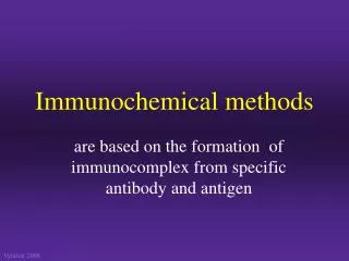
Applications of Immunochemical Methods in the Clinical Laboratory Roger L. Bertholf, Ph.D. Associate Professor of Pathology University of Florida College of Medicine
Particle methods Precipitation Immunodiffusion Immunoelectrophoresis Light scattering Nephelometry Turbidimetry Label methods Non-competitive One-site Two-site Competitive Heterogeneous Homogeneous Classification of immunochemical methods
Analytical methods using labeled antigens/antibodies • What is the function of the label? • To provide a means by which the free antigens, or antigen/antibody complexes can be detected • The label does not necessarily distinguish between free and bound antigens
Types of labels • Radioactive • Enzyme • Fluorescent • Chemiluminescent
Competitive Antigen excess Usually involves labeled competing antigen RIA is the prototype Non-competitive Antibody excess Usually involves secondary labeled antibody ELISA is the prototype Heterogeneous immunoassays
The birth of immunoassay • Rosalyn Yalow and Solomon Berson developed the first radioimmunoassay in 1957
Labeled antigen Specimen Wash Coated tube methods
Substrate E E E E E E 2nd antibody Specimen S P Microtiter well Enzyme-linked immunosorbent assay
E E E Labeled antibody S P Microparticle enzyme immunoassay (MEIA) Glass fiber matrix
Fe Fe Fe Fe Fe Fe Fe Fe Fe Magnetic separation methods
Fe Fe Fe Fe Fe Aspirate/Wash Magnetic separation methods
Oxidized Fe Reduced Electrochemiluminescence immunoassay(Elecsys™ system) Flow cell
Wash ASCEND
Developer ASCEND
Homogeneous immunoassays • Virtually all homogeneous immunoassays are one-site • Virtually all homogeneous immunoassays are competitive • Virtually all homogeneous immunoassays are designed for small antigens • Therapeutic/abused drugs • Steroid/peptide hormones
No signal Signal Typical design of a homogeneous immunoassay
Enzyme-multiplied immunoassay technique (EMIT™) • Developed by Syva Corporation (Palo Alto, CA) in 1970s--now owned by Behring Diagnostics • Offered an alternative to RIA or HPLC for measuring therapeutic drugs • Sparked the widespread use of TDM • Adaptable to virtually any chemistry analyzer • Has both quantitative (TDM) and qualitative (DAU) applications; forensic drug testing is the most common use of the EMIT methods
S S No signal S P Enzyme Enzyme Signal EMIT™ method
Functional concentration range EMIT™ signal/concentration curve Signal (enzyme activity) Antigen concentration
Fluorescence polarization immunoassay (FPIA) • Developed by Abbott Diagnostics, about the same time as the EMIT was developed by Syva • Like the EMIT, the first applications were for therapeutic drugs • Currently the most widely used method for TDM • Requires an Abbott instrument
Singlet E4 E3 E2 Triplet VR E1 IC A F 10-6-10-9 sec P 10-4-10 sec E0 Molecular electronic energy transitions
z x Polarizing filter y Polarized radiation
in out (10-6-10-9 sec) Fluorescein Fluorescence polarization Orientation of polarized radiation is maintained!
H O O O H O C O in out (10-6-10-9 sec) Rotational frequency 1010 sec-1 Fluorescence polarization But. . . Orientation of polarized radiation is NOT maintained!
Slow rotation Polarization maintained Rapid rotation Polarization lost Fluorescence polarization immunoassay
Functional concentration range FPIA signal/concentration curve Signal (I/I) Antigen concentration
Cloned enzyme donor immunoassay (CEDIA™) • Developed by Microgenics in 1980s (purchased by BMC, then divested by Roche) • Both TDM and DAU applications are available • Adaptable to any chemistry analyzer • Currently trails EMIT and FPIA applications in market penetration
Spontaneous Donor Acceptor Active tetramer Cloned enzyme donor Monomer (inactive)
Active enzyme No activity Donor Donor Acceptor Acceptor Cloned enzyme donor immunoassay
S S Fluorescence No signal Enzyme Enzyme S Signal Substrate-labeled fluorescence immunoassay
No signal Signal Fluorescence excitation transfer immunoassay
Oxidized Reduced Electrochemical differential polarographic immunoassay
P S P No signal Enzyme Enzyme P Signal Prosthetic group immunoassay
Substrate Product 1 E1 Product 2 E2 Enzyme channeling immunoassay
Early theories of antibody formation • Paul Ehrlich (1854-1915) proposed that antigen combined with pre-existing side-chains on cell surfaces. • Ehrlich’s theory was the basis for the “genetic theory” of antibody specificity.
The “Template” theory of antibody formation • Karl Landsteiner (1868-1943) was most famous for his discovery of the A/B/O blood groups and the Rh factor. • Established that antigenic specificity was based on recognition of specific molecular structures; he called these “haptens”; formed the basis for the “template” theory of antibody formation.
History of molecular imprinting • Linus Pauling (1901-1994) first suggested the possibility of artificial antibodies in 1940 • Imparted antigen specificity on native globulin by denaturation and incubation with antigen.
OH NH3+ N H 2 C l N O O O- O- Fundamentals of antigen/antibody interaction CH2-CH2-CH2-CH3
O O H H C C N N H H 3 3 N N N N O O N N CH3 CH3 Molecular imprinting (Step 1) Methacrylic acid + Porogen
O O H H C C N N H H 3 3 N N N N O O N N CH3 CH3 Molecular imprinting (Step 2)
O O H H C C N N H H 3 3 N N N N O O N N CH3 CH3 Molecular imprinting (Step 3) Cross-linking monomer Initiating reagent
In vivo preparation Limited stability Variable specificity General applicability In vitro preparation Unlimited stability Predictable specificity Limited applicability Comparison of MIPs and antibodies Antibodies MIPs
Immunoassays using MIPs • Therapeutic Drugs: Theophylline, Diazepam, Morphine, Propranolol, Yohimbine (2-adrenoceptor antagonist) • Hormones: Cortisol, Corticosterone • Neuropeptides: Leu5-enkephalin • Other: Atrazine, Methyl--glucoside