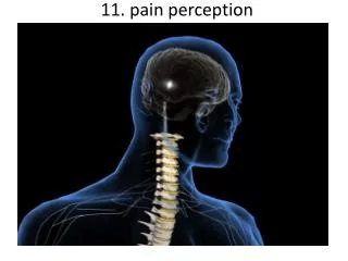
The perception of pain
540 likes | 1.36k Vues
The perception of pain. From Ch. 24 “Principles of Neural Science”, 4 th Ed. Kandel et al. Somatic sensations. Somatic sensation = bodily sensation Pain is a submodality of somatic sensation Pain and nociception (conscious vs. peripheral) Pain sensation is the most salient sensation

The perception of pain
E N D
Presentation Transcript
The perception of pain From Ch. 24 “Principles of Neural Science”, 4th Ed. Kandel et al
Somatic sensations • Somatic sensation = bodily sensation • Pain is a submodality of somatic sensation • Pain and nociception (conscious vs. peripheral) • Pain sensation is the most salient sensation • Pricking • Burning • Aching • Stinging • Soreness • Pain is a warning of actual or potential injury and damage • Pain depends on the psychological state • The same stimulus can result in different responses under similar conditions and in different individuals (soldiers, athlete, etc.)
Nociceptors • Information about stimuli that can damage tissue are conveyed by nociceptors • Chemicals are released from traumatized tissue • E.g. Substance P, histamine, and bradykinin • 3 classes of nociceptors • Mechnical: pinch, punctate, squeeze • Thermal: above 45 deg or below 5 deg Polymodal: mechanical, thermal, chemical Mechanical nociceptor
Somatic receptor types 1 2 3 4
Afferent fibers • Different size and conduction velocity of axons • Large fibers conduct faster than small/ thin fibers because the internal resistance to current flow is low and nodes of Ranvier are spaced further apart • Myelination sheets increase conduction velocity Compound AP = sum of all activated nerves Spike amplitude is proportional to fiber diameter
DRG Spinal dorsal horn Nociceptive afferents Compound Action Potential First pain: Sharp and pricking, faster A-delta fibers Second pain, burning and dull, slower C-fibers Blocking each nerve blocks the sensation
Spinal dorsal horn neurons • 2 overall types of interneurons: • Nociceptive specific: responds exclusively to noxious stimuli • Wide dynamic range neurons: graded response to non-noxious and noxious stimuli • Lamina I and II • Direct input from mainly A-delta and C fibers. • NS and WDR interneurons • Lamina III and IV • Direct input from A-beta. Nonnoxious input. Topographically organised receptive field • Lamina V • Direct input from A-beta and A-delta. Direct/ indirect from C-fibers. Convergence of visceral afferents. WDR interneurons projecting to brain stem and thalamus. • Lamina VI • Direct input from A-alpha (nonnoxious) from joints and muscle • Lamina VII and VIII • Respond to noxious input. Polysynaptic. Bilateral response
Neurotransmitters • Fast synaptic potentials • Glutamate (amino acid) • Efficient reuptake of amino acids • Range: postsynaptic neurons in vicinity • Slow synaptic potentials • Neuropeptides e.g. Substance P • No reuptake mechanisms • Range: diffusion, many neurons, unlocalized nature of pain • Neuropeptides • Released and increased in persistent pain conditions • Enhances and prolong the actions of glutamate • Application of substance P produces signs of inflammation e.g. heat, redness, and swelling
Chronic pain • Chronic pain appears to serve no useful purpose • Abnormal pain states • Nociceptive and neuropathic • Nociceptive pain • Direct activation of nociceptors • Tissue damage or inflammation • Neuropathic pain • Direct injury to the nerves • Peripheral or central • Burning or electrical sensation
Chronic pain • Spontaneous ongoing pain • Pain of variable intensity and duration • Spontaneous discharges in periphery and centrally • Referred pain • Pain in a location distant from the source. Could be explained by viscero-somatic convergence in lamina V • Hyperalgesia • Increased pain sensitivity • Allodynia • Non-painful input becomes painful e.g. touch on sun burned skin • Allodynia and hyperalgesia only exist during stimulation • Alterations in biochemical properties and excitability of dorsal horn neurons can induce spontaneous pain, hyperalgesia and allodynia
Referred pain • Signals from muscles and viscera can be felt as pain elsewhere • Example: myocardial infarction and angina can be felt in chest and left arm • Mechanism: convergence of afferents muscle/ viscera afferents and somatic afferents. • Convergence on the same projection neurons in the dorsal horn • The brain cannot tell the difference
Hyperalgesia • Peripheral sensitization: • Increased nociceptor sensitivity • Increased spontaneous activity • Central sensitization: • Increased spontaneous activity • Hyperexcitability of spinal dorsal horn neurons • Wind-up: progressive increased response = amplification (depends on glutamate acting on NMDA receptors) • Prolonged after-discharges to afferent input • Expansion of peripheral receptive fields of central neurons • Can be induced by repetitive firing of nociceptive afferents • Primary hyperalgesia • Hyperalgesia in damaged area (within 5-10mm) • Peripheral sensitization • Secondary hyperalgesia: • Hyperalgesia in surrounding undamaged tissue (10-20mm). • Peripheral and central sensitization
Pressure pain thresholds P<0.001 PTS CTR P<0.001 Niddam et al. 2008 Clinical hyperalgesia Myofascial pain patients (PTS) vs. normal controls (CTR) Myofascial trigger points are hyperalgesic contractures in the muscle IMES stimulus-response curves
Pain and the brain • Pain is a subjective conscious experience. Pain does not exist without the brain • CNS inhibitory or facilitatory mechanisms are remarkable efficient in decreasing or amplifying the pain experience • Changes in CNS contributes to chronic pain (reorganization: biochemical, atrophy, functions) • A better understanding of endogenous pain modulatory systems may lead to new mechanism-based therapies and drug targets
Pain and the brain: modulation • Factors that can influence the pain experience • Top-down brain processes • Memories (previous experience) • Emotion • Cognition (attention/ distraction) • Mood (depression, anxiety) • Context (stress, anticipation/ expectation, placebo) • Endogenous pain control systems • Other factors • Genes • Pathological factors (structure, transmitters, receptors, transporters etc.) • Age, gender
Acute vs. chronic pain • Acute pain characteristics • Activation of peripheral receptors under normal conditions • Sensation of pain closely related to the duration of the stimulus • Chronic pain characteristics • Spontaneous ongoing pain • Peripheral sensitization (spontaneous resting activity and hyperexcitable receptors) • Central sensitization (prolonged peripheral input) • Lowered pain threshold (Hyperalgesia) • Non-nociceptive input becomes painful (allodynia) • Functional and structural changes in PNS and CNS • Segmental expansion of receptive fields • De novo synthesis of membrane proteins • Spouting of spinal terminals of afferent fibers • Formation of new synaptic contacts • Altered balance in descending influences
Acute vs. chronic pain • It is important to differentiate between: • Acute and chronic pain states • Different time horizons engage different emotional coping strategies • Chronic pain becomes maladaptive and is highly co-morbid with mood and anxiety disorders • Chronic pain induces CNS changes • Ongoing spontaneous chronic pain vs. perturbations of chronic pain (allodynia/ hyperalgesia) • Passive vs. active coping => medial vs. lateral brain regions?
PFC PFC PFC ACC ACC PCC PPC PPC Insula PCC PCC Thalamus Insula Insula Vermis Amygdala Vermis Neuroimaging of acute pain Muscle pain Cutaneous pain Visceral pain Chen et al. Tooth pain Lu et al., 2004 Lin et al. (preliminary) Niddam et al., 2002
Distinct ascending pathways • Dorsal column-medial lemniscal system • Touch and proprioception from limbs and trunk • Somtatotopically organized from spinal to cortical level • Ascends ipsilateral side • Cross over to contralateral side in medulla • Spinothalamic pathway • Spinal lamina I, V-VII • Pain and temperature from limbs and trunks • Cross over to contralateral side in spinal cord • Somtatotopically organized from spinal to cortical level Contralateral
Ascending pathways • 5 major ascending pathways • Spinothalamic: axons of nociceptive specific and WDR neurons from laminae I and V-VII; contralateral projection, ascends in anterolateral white matter • Spinoreticular:neurons in laminae VII and VIII; anterolateral ascend • Spinomesencephalic: neurons in laminae I and V; anterolateral ascend to PAG, and spinoparabrachial tract to PB, amygdala; pain affect • Cervicothalamic: arises from lateral cervical nucleus; laminae III and IV; some projects via the dorsal column to cuneate and gracile nuclei (large fiber pathway) • Spinohypothalamic; laminae I, V, VIII; autonomic control • Thalamic nuclei • Lateral nuclear group: spinothalamic tract, NS and WDR, laminae I and V, small receptive fields, encoding location of injury • Medial nuclear group: spinoreticulothalamic tract, laminae VII and VIII
Apkarian et al. 2005 Millan 2002 Pain pathways in the brain Spino-bulbo-spinal loop (pain facilitation) Ascending pathways and cortical/ sub-cortical connectivity Pain components (variable expression): Sensory-discriminative, affective-motivational Cognitive, Motor
Pain and the brain: pathways • Stress and the reward/ motivation system Dopamine based mesolimbic system modulates mainly tonic pain Hypothalamus Amygdala Hippocampus Ventral striatum/ Nucleus accumbens Ventral tegmental area Dopoaminergic nucleus Ventral pallidum MDm thalamus Anterior cingulate Pain modulation
Motivation and emotions Borsook 2007, EJP
Pain modulation: large fibers • The balance of activity in small- and large-diameter fibers is important in pain transmission/ determines the pain intensity • The gate control theory involves 4 types of neurons in the dorsal horn of the spinal cord • Large-diameter afferent (non-nociceptive) • Small-diameter afferent (nociceptive) • Inhibitory interneurons (spontaneously active) • Projection neurons • Large-diameter fibers • Excites interneuron and decrease pain transmission/ Closes the pain-gate • Small-diameter fibers • Inhibits interneuron and increases pain transmission/ Opens the pain-gate • Observation: in absence of conduction in A-ά/ A-β fibers pain perception is abnormal • Pin prick, pinch, ice cold produces burning pain The gate control hypothesis
Pain modulation: opiods • Direct stimulation of PAG produces analgesia • Inhibits firing of nociceptive neurons in lamina I and V • Descending pathway recruited: PAG excites rostroventral medulla/ nucleus raphe magnus (5HT) • Moprhine (an opioid) induced analgesia via endogenous opioid receptors in descending pathway • Opioid receptors • Types: -, δ-, κ-, nociceptin • Transmitters: enkephalins, β-endorphin, dynorphin • Location (mainly): PAG, ventral medulla, superficial dorsal horn • Stress-induced analgesia of escapable pain is mediated via the endogenous opioid system • Side effects • Other regions not involved in pain also contains opioid receptor • Minimize diffusion by local administration can avoid side effects e.g. in cerebrospinal fluid