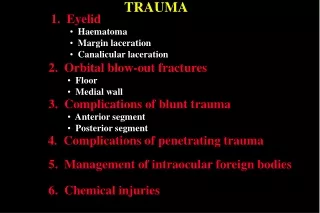
TRAUMA
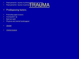
TRAUMA. Peak period for injuries in primary teeth are 2-4 years Peak period for injuries in permanent teeth 8-10 years Predisposing factors : Protruding upper incisors Incompetent lip Bad eye sight Physical and mental handicapped Gender Central incisors.

TRAUMA
E N D
Presentation Transcript
TRAUMA • Peak period for injuries in primary teeth are 2-4 years • Peak period for injuries in permanent teeth 8-10 years • Predisposing factors: • Protruding upper incisors • Incompetent lip • Bad eye sight • Physical and mental handicapped • Gender • Central incisors
Dental trauma is considered an emergency condition : relieve of pain and reduction of displaced teeth • Rational therapy depends upon correct diagnosis whereas incomplete examination leads to inaccurate diagnosis and less successful treatment.
History • 1. Patient name, age etc • 2. When did the occur? • Time factor influence the choice of treatment, (Implantation, P. exposure) • 3. Where did injury occur? • Tetanus prophylaxis, antibiotic, legal implication
History • 6. Medical history • 7. Subjective complaints: • a. Did trauma causes amnesia, unconsciousness. vomiting or • headache, indicate brain concussion. • b. Is there any disturbance in bite, affirmative answer may imply: • tooth luxation, alveolar fracture , jaw fracture, fracture of the TMJ. • c. Reaction to thermal or other stimuli.
Clinical examination :Provides all information necessary to make correct diagnosis and design an appropriate treatment plan • Before initiating the C.E. the areas should be cleaned of all debris and blood.
Clinical Examination should include • 3: Bony fracture • 4: Hemorrhage into floor of the mouth J Fra. • 5: Cracks and Craze lines, fracture, pulp exposure, discoloration of the crown.(Light) • 6: Displacement of the tooth in any direction
Clinical Examination should include: • 7: Abnormal, horizontal & vertical mobility • 8: Injury to PDL. • 9: Abnormalities in the occlusion. • 10: Percussion test • 11: Reaction of teeth to sensibility testing:
11: Reaction of teeth to sensibility testing: • 1. Heat, heated gutta percha • 2. Cold, ethyl chloride, carbon dioxide snow -78, dichlorodifluoromethane -28. • 3. Electric pulp tests: • Before initiating vitality testing, the following factors should be considered: • A. erupting non-injured teeth • B. testing should be conducted away from the gingival area • C. splinted and crowned teeth. • D. since pt. adapt sustained E C, pain threshold should be determined by rapid, steady increase in current rather than slow, gradual increase.
Radiographic Examination • Radiographic E. should reveal the following information: • 1. Root fracture • 2. Degree of intrusion or extrusion • 3. Presence of periapical rarefaction • 4. Extent of tooth development • 5. Size of the pulp chamber or root canal • 6. Foreign bodies lodged in the soft tissue
2. Fracture Line are usually obliquely positioned. 10-15 apical or coronal. • 3. A bend in a film Tracing P ligament around the root. • 4. After six week another x-ray.
Classification of Dental trauma • Modification of Ellis (1945) • Class 1 fracture enamel • Class 2 fracture enamel and dentine • Class 3 fracture E/D with pulp involvement • Class 4 discoloration of the crown • Class 5 displacement • Class 6 tooth lost • Class 7 tooth restored by composite or crown
Why it is important to restore crown anatomy: To prevent labial protrusion and displacement. To prevent drifting or tilting of the fractured tooth. To prevent over-eruption of opposing teeth. To protect the vitality of the tooth. To restore aesthetic .
A conventional two‑bottle adhesive system (Scotch bond Multi‑Purpose Plus, 3M ESPE applied on the enamel.( After juxtaposition)
Concussion and Subluxation • Concussed Tooth is tender to percussion(vertical & horizontal) due to edema and Hemorrhage in PDL • Subluxated Tooth is tender to percussion and abnormally loose due to rupture of PDL , gingival hemorrhage around the gingival margin • Treatment: • adjustment of occlusion (slight grinding of opposing tooth) • soft diet • splinting for the comfort of the pat. Does not prompt healing • repeated sensibility testing
Intrusion Luxation • Displacement of the tooth deeper into the alveolar bone with comminuting or fracture of the alveolar bone (axial force). Traumatic intrusion of permanent teeth is a rare injury, which represent up to 2% of all traumas affecting permanent dentitions. • Clinical findings: • the affected tooth appears shorter than the contra lateral tooth • high metallic sound in percussion • no mobility • x-ray: PL space partially or totally disappears • pulp necrosis 63% in immature teeth and 100% in mature ones.
Teeth with immature root development, spontaneous re-eruption should be awaited. Teeth with mature root development in patients in the age interval 12–17, spontaneous re-eruption could be awaited. In patients older than 17 with mature root formation, orthodontic or surgical repositioning should be performed.
avulsion • Maxillary incisors (prominent) • Children 7-10 years old • Success rate 4-50% • Two important aspects in the successful treatment: • A. the condition under which the tooth has been preserved. • B. the time interval between the injury and the treatment • The following condition to be considered before R.A.T: • A. The avulsed tooth should be without advanced P.diseas • B. The alveolar process should be intact to provide a seat • C. Their should be no orthodontic contraindication • D. The stage of root development should be evaluated
To provide the best chance of success, the PL cells should be kept in the most physiologically healthy status as possible. • If the avulsed tooth does not replanted within 60-120 min., the PL cells undergo necrosis because PL cells deprived from its blood supply and depletion of the stored cell metabolites, then root resorption begins and leads to the loss of pre-cementum layer.
Storage media • Water and saline are damaging to PL cells (one hour) • Milk (low fat) limited in benefit (two hours) • Hank’s Balance Saline Solution (HBSS) (Save-A-tooth sys) • Gingival fluid storage media not available commercially • Emdagon is an enamel matrix derivative gel Promote regeneration of PL cells. • egg white, isotonic solutions, propolis • and green tea, soymilk
Avulsion of primary teeth has been reported to comprise between 5.8% and 19.4% of all types of traumatic injuries to the primary. It occurs most often in 2–4-year-old children and affects boys 1.2–1.5 times more than girls. The maxillary primary central incisor is involved more than any other tooth followed by maxillary lateral incisors and mandibular central incisors . The main causes of avulsions of primary teeth are falls, fights and child abuse. Three options are possible for management of an avulsed primary incisor: (i) no treatment, avoid replantation), (ii) prosthetic replacement of the missing tooth and (iii) replantation of the avulsed tooth.
Complication of primary teeth injuries on developing permanent teeth • white or brown discoloration of the permanent tooth with or without hypoplastic defects; • dilaceration of the crown of the tooth causing eruption disturbance or failure . • dilaceration of the root of the tooth causing eruption disturbance or failure; odontome-like formation. • partial or total failure of root development; • total failure of tooth development .
Replantation of primary teeth should be avoided in the following cases: 1. When the crown of the permanent successor is not yet completely developed. 2. Children with systemic diseases. 3. Children with behavior disorders whose compliance with postoperative instructions is expected to be problematic. 4. Multiple avulsions (no teeth for splinting). 5. Severe fracture of the alveolar bone. 6. Tooth close to natural shedding. 7. Root resorption due to previous trauma. 8. Severely decayed teeth.