Download
1 / 43
550 likes | 1.43k Vues
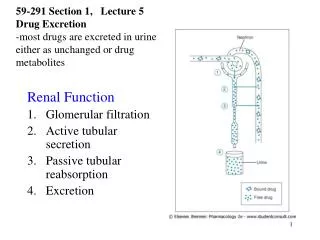
Tubular reabsorption and tubular secretion. Type of Transport. Transcellular reabsorption Primary active transport Secondary active transport Ion channel – passive transport Paracellular reabsorption. Tubular Reabsorption. LUMEN. apical. basal. BLOOD.

E N D
Type of Transport • Transcellular reabsorption • Primary active transport • Secondary active transport • Ion channel – passive transport • Paracellular reabsorption
LUMEN apical basal BLOOD
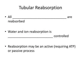
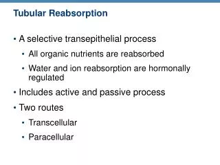
Reabsorption routes and transport mechanisms • Reabsorption routes 1- Paracellular reabsorption • Between adjacent tubule cells • Tight junction do not completely seal off interstitial fluid from tubule fluid • Passive 2- Transcellular reabsorption – through an individual cell • Primary active transport (ATP) • Sodium-potassium pumps in basolateral membrane only • Secondary active transport (co-transport) • Symporters, Antiporters (exchange) • Transport maximum (Tm) • Upper limit to transport a substance • Obligatory (Osmosis) vs. facultative (ADH) water reabsorption
Mechanisms of secondary active transport
Sodium reabsorption in proximal convoluted tubule (PCT) 65 -70% of filtered sodium is reabsorbed in PCT Followed by water & chloride Sodium is important for the absorption of Glucose Amino acids phosphates
NephronTubular Reabsorption Na+ reabsorption An active process • Occurs on the basolateral membrane (Na+/K+ ATPase) • Na+ is pumped into the interstitial fluid • K+ is pumped into the tubular cell • Creates a Na+ gradient that can be utilized for 2ºry active transport
NephronTubular Reabsorption Secondary Active Transport utilizing Na+ gradient (Sodium Symport) Used for transporting: Glucose, amino acids, ions, metabolites
Mechanism of Na Reabsorption Steps • Luminal side. Movement of Na from lumen to intracellular fluid by electrochemical gradient. • Basolateral membrane, Na+/K+ ATPase, 3 Na / 2 K pumped, results in Low intracellular Na concentration and osmolarity in the basolateral space. • Peritubular side (bulk flow). Movement of hyperosmotic fluid from basolateral spaceto peritubularcapillaries.
H + Na + glucose, amino acids Na + Urea H20 Cl- Reabsorption of Water and Solutes is Coupled to Na+ Reabsorption Tubular Cells Tubular Lumen Interstitial Fluid - 70 mV Na + ATP K+ Na + ATP Na + K+ 0 mv - 3 mV
Movement of NaCl & water into peritubular capillaries (Bulk Flow) NaCl enter the peritubular capillaries by simple diffusion Water follows Na due to the high oncotic pressure in the capillaries In peritubular capillaries the high plasma oncotic pressureis due to fluid filtration in glomerulus GFR oncotic pressure & hydrostatic pressure in efferent & peritubular capillaries bulk flow from lateral space to peritubular capillaries reabsorption GFR oncotic pressure & hydrostatic pressure bulk flow fluid go back to lumen through tight junction reabsorption
Mechanisms by which Water, Chloride, and Urea Reabsorption are Coupled withSodium Reabsorption
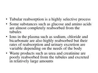
Water reabsorption 60-70 % of filtered water is reabsorped in PCT Active transport of Na from the renal cell to peritubular space Increased osmolality of peritubular space Drag water by osmosis Filterate remain iso-osmotic (~equal quantity of water & solute are absorbed)
TUBULAR REABSORPTION OF GLUCOSE, AMINO ACIDS, UREA & OTHER ELECTROLYTES
Glucose reabsorption general consideration • Glucose reabsorption is calculated as the difference between the amount of glucose filtered by the kidney and the amount excreted. • When plasma glucose (PG) is increased to near 200 mg/dl, glucose begins to appear in urine – this is called the “glucose renal threshold” corresponds to filtered load of 250mg/min.
As glucose is further increased, more glucose appears in urine. • At very high filtered glucose, reabsorption remains constant, this is called “tubular transport maximum” for glucose (TmG) • = 375 mg/min (corresponds to blood glucose level of 300mg/dl) At this maximum transport, all glucose carriers are saturated and no more glucose can be transported.
K+ Na+ Glucose Glucose GLUT 2 SGLT 2 One Na+ Early proximal tubule cell Glucose Reabsorption INTERSTITIAL FLUID TUBULAR LUMEN K+ Na+ Glucose Glucose GLUT 1 SGLT 1 Two Na+ Late proximal tubule cell
Tubular maximum (TmG) Maximum absorptive capacity for glucose by renal tubular cells = 375 mg/min in males (females 300mg/min) • Renal threshold Plasma glucose level at which glucose first appear in urine = 200mg/dl in arterial; 180 mg/dl in venousblood.
Amino acid reabsorption • All filtered aminoacidsare reabsorbed in PCT • Luminal membrane Cotransport with Na • Basolateralmembrane diffusion
Bicarbonate reabsorption • 90% of filtered bicarbonate is reabsorbed in PCT • Filtered HCO3 + H H2CO3 • H2CO3 H2O + CO2 • CO2diffuses into the cell + H2O H2CO3 in the presence of carbonic anhydrase • H2CO3 H + HCO3 • HCO3is reabsorped • H+ is secreted in exchange for Na +
Bicarbonate reabsorptioncont. Lumen Tubular cell Blood Filtrate Na+ HCO3 + H+ H2CO3 H2O + CO2 Na+ H+ HCO3 H2CO3 CA CO2 + H2O Tight junction Brush border
Phosphate reabsorption • Freely filtered • 1/3 of filtered load is excreted in urine • Cotransportedwith Na • Compete with glucose: blocking glucose Phosphate reabsorption
Urea reabsorption • Plasma urea concentraion 15-40mg/100ml • End product of protein metabolism • 40-50% of filtered urea reabsorbed • Passive diffusion • Coupled with Na reabsorption • 50-60% excreted
Urea reabsorption cont. • GFR (renal disease; low renal blood flow) urea concentraion in plasma • GFR urea filtered • GFR slow flow rate of filterate more urea is absorbed to blood
Filtration, reabsorption, and excretion rates of substancesby the kidneys Filtered Reabsorbed Excreted Reabsorbed (meq/24h) (meq/24h) (meq/24h) (%) Glucose (g/day) 180 180 0 100 Bicarbonate (meq/day) 4,320 4,318 2 > 99.9 Sodium (meq/day) 25,560 25,410 150 99.4 Chloride (meq/day) 19,440 19,260 180 99.1 Water (l/day) 169 167.5 1.5 99.1 Urea (g/day) 48 24 24 50 Creatinine (g/day) 1.8 0 1.8 0
Tubular secretion • Potassium: 90% of filtered K is reabsorbed (PCT) K secreted in DCT In exchange for Na; under the control of Aldosterone • Hydrogen: Excretion is inversely proportional to K
Proximal Tubule GFR delivers ~125ml/min of filtered plasma to the nephron (tubule) • Nearly 70% of this volume is reabsorbed by the proximal tubule and returned back to the blood. • Needed products such as glucose and amino acids are reabsorbed rapidly whereas; • Other products such as creatinine and urea are slowly reabsorbed or not at all • Proximal tubule also secretes substances that the body need to get rid of rapidly. These include toxins, drugs, H+, urea and ammonia.
Proximal tubule transports the largest %age of most substances of the entire nephron. • about 70% of the filtered H2O is reabsorbed in the proximal tubule • About 70% of the filtered Na+ is reabsorbed in the proximal tubule • Therefore ~30% of the fluid filtered (GFR) is delivered to the “Loop of Henle”
Cellular Ultrastructure and Primary Transport Characteristics of Proximal Tubule
The Loop of Henle Thin descending loop Major function → H2O reabsorption • At start of descending loop osmolarity is same as plasma (~280 mOsm) • At end of descending loop osmolarity = 1200 mOsm • Most of the osmolarity is due to NaCl and little due to Urea
Cellular Ultrastructure and Transport Characteristics of Thin and Thick Loop of Henle very permeable to H2O) not permeable to H2O)
NaCl Urea NaCl Urea 280 mOsmol/kg H2O H2O H2O H2O H2O NaCl Urea NaCl Urea 1200 mOsmol/kg H2O
Thin & thick ascending loop • No H2O reabsorption • Increases solute reabsorption (mainly NaCl) • This part of the loop (thin ascending loop) is very permeable to Na+ and Cl¯ • Passive diffusion out of tubule • As NaCl diffuses out of the tubule, the osmolarity becomes more dilute due to the movement of only solutes and not H2O
In thick ascending limb reabsorption of NaCl is active (requiring energy). Movement of NaCl from tubule to interstitial fluid is active • Na+-K+-2Cl¯cotransport causes movement of these ions into cells from tubule lumen • Na+ actively transported out of cell • Cl¯ increases in cell due to the cotransporter (Na+-K+-2Cl¯cotransport). This causes an increase in Cl¯ in the cell which is greater than the interstitial fluid. Therefore Cl¯ moves out of cell passively.
Tubular lumen Tubular Epithelial Cells Renal interstitium Na+ 3Na+ Na+ 2K+ 2Cl¯ 2Cl K+ K+ +10-20 mV
Distal Tubule and Collecting Duct • Function as proximal tubule but absorbs less and has a smaller capacity: • Reabsorbs 9% filtered Na+ • Reabsorbs 19% filtered H2O • Thoughout distal tubule Na+is actively reabsorbed and K+secreted • Cl¯ also reabsorbed
Reabsorption and secretion in the late distal convoluted tubule and collecting duct • 90-95% of filtered solutes and fluid have been returned by now • Principal cells reabsorb Na+ and secrete K+ • Intercalated cells reabsorb K+ and HCO3- and secrete H+ • Amount of water reabsorption and solute reabsorption and secretion depends on body’s needs
Cellular Ultrastructure and Transport Characteristics of Early and Late Distal Tubules and Collecting Tubules Not permeable to H2O Not very permeable to urea permeability to H2O depends on ADH not very permeable to urea
Hormonal regulation of tubular reabsorption and secretion • Angiotensin II - when blood volume and blood pressure decrease • Decreases GFR, enhances reabsorption of Na+, Cl- and water in PCT • Aldosterone - when blood volume and blood pressure decrease • Stimulates principal cells in collecting duct to reabsorb more Na+ and Cl- and secrete more K+ • Parathyroid hormone • Stimulates cells in DCT to reabsorb more Ca2+
Regulation of facultative water reabsorption by ADH • Antidiuretic hormone (ADH or vasopressin) Increases water permeability of cells by inserting aquaporin-2 in last part of DCT and collecting duct • Atrial natriuretic peptide (ANP) • Large increase in blood volume promotes release of ANP • Decreases blood volume and pressure by inhibiting reabsorption of Na+ and water in PCT and collecting duct, suppress secretion of ADH and aldosterone