Download

1 / 46
460 likes | 484 Vues
Apoptotic Cell Clearance and the Resolution of Inflammation. Jeremy Hughes MD PhD Wellcome Trust Senior Research Fellow in Clinical Science MRC Centre for Inflammation Research, University of Edinburgh. Talk Outline: Apoptosis in inflammation Current apoptotic cell recognition mechanisms

E N D
Apoptotic Cell Clearance and the Resolution of Inflammation Jeremy Hughes MD PhD Wellcome Trust Senior Research Fellow in Clinical Science MRC Centre for Inflammation Research, University of Edinburgh.
Talk Outline: • Apoptosis in inflammation • Current apoptotic cell recognition mechanisms • Regulation of macrophage phenotype by apoptotic cell ingestion • Efficient apoptotic cell clearance limits autoimmune responses
Physiological apoptosis • Embryological development • Tissue homeostasis • Regulation of leukocyte populations e.g. neutrophils • Deletion of autoreactive T cells in thymus • Pathological apoptosis • Inflammation • Infection • Cancer • Autoimmunity
Renal Cell Apoptosis - TUNEL staining Proximal tubule Interstitial cell
Elevated levels of apoptosis documented in disease states/experimental models e.g. • Obstructive nephropathy • Gobe ands Axelsen demonstrated that excess apoptosis resulted in tubular atrophy (Lab Invest. 1987 56:273) • Mesangial proliferative glomerulonephritisBaker et al demonstrated that mesangial cell apoptosis is critically important in resolving Thy 1 GN(JCI 1994 94:2105)
Large scale renal cell apoptosis is pro-inflammatory • Ischaemia reperfusion injury (kidney/cardiac) • (Daemen et al, University of Maastricht) MIP-2 and KC levels APOPTOSIS Renal Dysfunction Neutrophil infiltrate
Anti-apoptosis treatments are protective • ZVAD • IGF-1 • Acute phase proteins MIP-2 and KC levels APOPTOSIS Renal Dysfunction Neutrophil infiltrate
Apoptosis May Be Pro-inflammatory Apoptosis Excess death or Defective Phagocytosis Rapid efficient phagocytosis Pro-inflammatory (MCP-1, IL-8 from apoptotic cell) Anti-inflammatory
Pro-inflammatory • Tissue atrophy APOPTOSIS = double edged sword • Resolution of hypercellularity • Modulation of Mø function
An Intraperitoneal Competition Assay Apoptotic cells • Injected IP • 30 min incubation • Mø rich greater omental lymphoid organ excised Live cells
Preferential and rapid clearance of apoptotic cells Cell Number per field 300 200 100 0
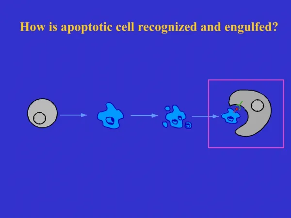
The ‘phagocytic synapse’ Savill et al Nature Rev 2002 2:965
Multiple receptor families involved • Phagocyte surface receptors: • Integrins (b1, b2, b3, b5) - Adhesion molecules and ECM • Scavenger receptors (SRA, CD36) - Lipids • Complement receptors - Pathogens • CD14 - bacterial lipopolysaccharide (LPS) • Phosphatidylserine receptor (PSR) • Lectins
Multiple receptor families involved • Bridging molecules: • Thrombospondin • C1q • Apoptotic surface: • Phosphatidylserine (PS) • ICAM-3 • Sugars • ‘Apoptotic cell associated molecular patterns’
Phagocyte receptors involved are multifunctional CD14 + LPS NFkB activation TNFa secretion
Phagocyte receptors involved are multifunctional CD14 + Apoptotic cell Increased TGF decreased NFkB No TNFa secretion CD14 + LPS NFkB activation TNFa secretion ‘Turn off’ signal
Apoptotic cell clearance regulates macrophage phenotype and promotes the resolution of inflammation
Classically vs Alternatively Activated Macrophages • ‘Angry’ macrophage • TNFa & cytokines • Nitric oxide (NO) • ROS • ‘Healing’ macrophage • IGF1 • PDGF • bFGF • VEGF • TGFb • Classical Activation: • Pathogens (LPS, DNA) • IFNg and TNFa, IL-1 • Alternative Activation: • IL-4, IL-10, IL-13 • TGFb • Glucorticoids
Macrophages in Inflammation and Tissue Healing • Resolving Inflammation • Alternative Activation predominates: • Anti-inflammatory mediators • Pro-cell survival • Pro-angiogenesis • ECM stabilisation • Acute Inflammation • Classical Activation predominates: • Pro-inflammatory mediators • Cell killing • Pathogen killing • ECM degradation
Interaction with apoptotic cells exerts critical effects upon macrophages
Macrophage Response to Apoptotic Cell Ingestion • Macrophage release of autocrine and paracrine mediators: • TGFb • PGE2 • PAF • Downregulated expression of ‘killer molecules’: • iNOS • TNFa
Macrophage Response to Apoptotic Cell Ingestion • Net effect is: • ‘deactivation’ of macrophages and • ‘re-programming’ to reparative phenotype • AC ingestion also: • increases macrophage survival and • reduces macrophage proliferation • (Reddy et al. J Immunol 2002 169:702)
4. MC induced into apoptosis 5. Phagocytosis inhibits further killing TNF/NO 3. killing 1. Activated Mø 2. Susceptible mesangial cell
Phagocytosis adds ‘new meaning’ to cell death Savill et al Nature Rev 2002 2:965
Can we harness the potential power of the interaction of macrophages with apoptotic cells?
AC Administration Ameliorates Lung Injury PBS Apoptotic Cells d1 d3 Huynh et al JCI 2002 109(1):41
Corticosteroids: • Induce lymphocyte apoptosis • Sensitise mesangial cells to apoptosis • Significantly increase macrophage capacity to ingest apoptotic cells • AC clearance upregulated by: • Cytokines • Lipoxins
Efficient apoptotic cell clearance prevents the generation of autoimmune responses
Apoptotic cells contain potential autoantigens Normal Cell Apoptotic Cell
Apoptosis and Autoimmunity Apoptosis Inadequate Mø/resident cell Phagocytosis Excessive level of apoptosis Apoptotic cell ingested by dendritic cell and potential antigen presentation Autoimmune response
C1q and Apoptotic Cell Clearance Apoptotic Cells + C1q WT serum • Injected IP into C1q KO mouse • 30 min incubation • Greater omental lymphoid organ excised Apoptotic Cells + C1q KO serum
C1q opsonisation augments apoptotic cell clearance Cell Number per field
Clq and autoimmunity • Clq deficient patients have high incidence of developing SLE • Clq KO mice spontaneously develop autoimmune glomerulonephritis with excess apoptotic cells in the kidney • Clq KO mice develop more severe NTN
Organ and Cell Specific Subtleties in Apoptotic Cell Clearance
PMNs - specific upregulation of apoptotic PMN clearance by Ab ligation of macrophage CD44 • Lungs - surfactants involved in apoptotic cell clearance • ClqKO mice - • excess apoptotic cells evident in kidney • defective clearance of AC in the peritoneum • normal AC clearance in UV irradiated skin
Clinical Implications • Modulation of apoptotic cell clearance may provide novel treatments for diseases characterised by: • Acute inflammation and marked cell death • Macrophage dependent tissue injury • Autoimmune responses
Acknowledgements EdinburghFunding Tiina Kipari Wellcome Trust Simon Watson Medical Research Jean Francois Cailhier Council Claire Taylor Michael Clay Kris Houlberg