Download
1 / 25
270 likes | 547 Vues
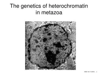
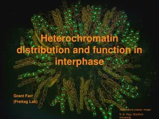
Heterochromatin distribution and function in interphase. Grant Farr (Freitag Lab). Neurospora crassa image: N. B. Raju, Stanford University. Long-term objectives. To find fungus-specific inhibitors to better combat fungal infections in humans, animals and plants.

E N D
Heterochromatin distribution and function ininterphase Grant Farr (Freitag Lab) Neurospora crassa image: N. B. Raju, Stanford University
Long-term objectives • To find fungus-specific inhibitors to better combat fungal infections in humans, animals and plants. • To determine if centromere and heterochromatin organization are involved in polarized growth. Aspergillosis X-Ray by ADAMS Health Care Center
Vegetative cycle Histone H1-GFP tagged nuclei M. Springer Neurospora crassa
Polarized hyphal growth Histone H1-GFP tagged nuclei
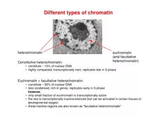
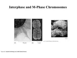
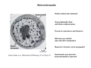
Chromatin is heterogeneous euchromatindecondensed, active, DNA unmethylatedhistones hyperacetylated heterochromatincondensed, inactive, DNA methylatedhistones hypoacetylated Arabidopsis fission yeast mouse Wolffe (1998) Chromatin
Nuclear architecture Chromocenter Region of heterochromatin that may be associated with the telomeres HP1-GFP Histone H1-GFP
Role of heterochromatin in polarized growth HP1 mutants exhibit slow linear growth
Centromere-specific proteins CENP-A (CenH3) Centromere-specific histone H3 Other proteins: ~30 proteins (CENP-B to CENP-S) in humans ~60 known proteins in yeast Neurospora shares four identifiable proteins with humans: CenH3 Cenp-I Cenp-S CAC-3 (CAFp46/48) Young-Tae Chang and Young-Soo Kim from NYU department of Chemistry
DH5 DH5 DH5 Experimental outline 1. PCR 2. Digest plasmid and insert 3. Ligate 5’ 3’ XbaI BamHI 3. Transformation into competent E.coli pMF272 pGF1 6. Analysis by epifluorescent microscopy 4. Purify DNA, linearize 5. Transform N. crassa
Amplification of genes for centromere-specific proteins + all three genes were amplified successfully + Cenp-S was fused to GFP + Cenp-I was fused to RFP + CAC-3 fusions did not work M CENP-S CENP-I CAC-3 3.0 kb 2.0 kb 1.0 kb 0.5 kb CCG1 promoter RFP Cenp-I C-Terminus CCG1 promoter Cenp-S GFP C-Terminus
Transformation of Neurospora crassa • Transformed linearized plasmid DNA carrying Cenp-S and Cenp-I fusion genes into two N. crassa strains each • Genes targeted to the his-3 locus Initial transformations of Cenp-S Initial transformation of CENP-I
Backcross to purify strains Uncrossed CENP-I RFP Crossed CENP-I with expected localization and less background noise A.J.F. Griffiths, U.B.C. • Crosses: Cenp-S-GFP X N2557 (ridhis-3 mat a) Cenp-S-GFP X N2556 (his-3 mat a; hpoRIP2) RFP-Cenp-I X N2557 (ridhis-3 mat a) RFP-Cenp-I X N2556 (his-3 mat a; hpoRIP2)
The HP1 cells previously characterized have similar localization. CenH3 localization is identical to Cenp-S localization. Cenp-S is identified as a part of the centromeric Cenp-A complex. Cenp-S labels the centromere HP1 labeled with centromere and telomeric regions fluorescing Cenp-S-GFP
Imaging of the Labeled Centromeres Cenp-S gfp localized at the centromere
Imaging of the Labeled Centromeres Cenp-I localized in the centromeric DNA region.
Confirming centromeric localization Patrick Hickey (University of Edinburgh) • Hypha of different strains can form heterokaryons. • Fusing SON-1-GFP and HP1-GFP strains with the new Cenp-S and Cenp-I strains will show simultaneous localization of the nuclear membrane and centromeres.
Cenp-I and HP1 co-localize partially HP1-GFP RFP-CENP-I
SON-1-GFP + RFP-CENP-I • Cenp-I localization is centromeric SON-1 gfp CENP-I rfp
Is HP1 required for centromere localization? • Hypothesis: Heterochromatin is required for centromere localization. • I tested this with the hpo X Cenp-S and Cenp-I crosses.
Localization of CENP-S and CENP-I is maintained in HP1 mutants • The same appears to be true for CenH3 (data not shown) • Centromere localization appears independent of heterochromatin. CENP-S-GFP; hpo RFP-CENP-I; hpo
Possible Cenp-I mutant • One of the Cenp-I X wildtype crosses gave us an hpo mutant like growth phenotype. • The phenotypes are separable by microscopy. • In the future we will sequence the Cenp-I genes to search for point mutations introduced by RIP in the cross.
Summary • The Cenp-I and Cenp-S fusion proteins were expressed and appear to be located at centromeres. • Lack of heterochromatin binding protein does not affect the localization of either proteins. • Possible mutant of Cenp-I affects apical growth.
Future Studies • Construct an RFP-Cenp-S strain and a Cenp-I-GFP strains to better understand localization of the two using heterokaryons. • Slow-growing strains will be tested for the hpo mutation by DNA sequencing to verify that Cenp-S and Cenp-I localization were independent of HP1. • Use protein affinity tags (TAP, Myc, HA) to characterize other proteins in centromere complexes. • Find mutants in the Neurospora “knockout” collection that affect centromere localization and polarized growth.
Acknowledgements • Howard Hughes Medical Institute • Dr. Kevin Ahern • Dr. Michael Freitag • Thomas Lew