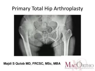
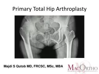
Primary Total Hip Arthroplasty
Primary Total Hip Arthroplasty. Majdi S Qutob MD, FRCSC, MSc, MBA. History. 1891 Themistocles Glück (1853-1942) carried out the first reported Femoral Hemiarthroplasty in Germany, using ivory to replace the femoral head

Primary Total Hip Arthroplasty
E N D
Presentation Transcript
PrimaryTotal Hip Arthroplasty Majdi S Qutob MD, FRCSC, MSc, MBA
History • 1891 Themistocles Glück (1853-1942) carried out the first reported Femoral Hemiarthroplasty in Germany, using ivory to replace the femoral head • 1940 Dr. Austin T. Moore (1899–1963), first metallic (Vitallium) Femoral Hemiarthroplasty • 1960 Dr. San Baw (1922 –1984) ivory Femoral Hemiarthroplasty for neck fractures • 1970 Dr Sir John Charnley (1911 –1982) Low Friction Arthroplasty
Demographics By 2030, the demand for primary total hip arthroplasties is estimated to grow by 174% to 572,000 The Journal of Bone & Joint Surgery. 2007; 89:780-785
Arthritis • Arthritis is the second most common chronic condition in the US (sinusitis is first) • Most common among elderly • 20-30% of people over age 70 suffer from osteoarthritis (OA) of the hip • Arthritis affects over 32 million people in the US • Total costs associated with arthritis are over $82B/year, including hospital and drug costs, nursing home costs, and lost productivity and work
Normal ROM IR- 35° ER - 45° Flexion - 135° Extension - 15° Abduction - 45° Adduction - 25°
Non-Surgical Intervention • Activity Modification • Weight Loss • Cane/walker • Physical Therapy • Medications: • NSAIDs • COX-2 Inhibitors • Nutritional supplements • Injections: • Corticosteroid • Viscosupplementation
Joint Anatomy • Femoral Head Diameter 46mm • Neck Shaft Angle Average 130 degrees • > 135 degcoxavalga • < 120 deg, coxavara • Femoral Anteversion Angle 12-15 degrees • Femoral Offset variable • Acetabular Anteversion Average 15 (0-20) degrees • Acetabular Abduction Angle 45 degrees (30-50) • AP Curve of Femur is about 4 degrees
Biomechanical Considerations • 1-2million steps/year • 3-6x body weight due to abductors- 7-8x sporting activities • Descending stairs causes highest JRF • Abductors provide two thirds of the hip joint force parallel to the long axis of femur • Increasing the offset and cup medialization reducing the joint reaction force by increasing the abductor lever arm
Biomechanical Considerations • Increasing the offset and cup medialization reducing the joint reaction force by increasing the abductor lever arm • 10mm increase femoral offset decreases 10% abductors force requirement
Bone Biomechanics • Young’s Modulus (flexibility) =stress/strain • Stress =force/area • Strain= change in length • Bone is anisotropic (compression>shear) • Exhibits creep: constant pressure will deform at a decreasing rate • Strain rate low with rapid application of modest force leads to fracture
Singh Classification for Femoral Neck Osteoprosis Singh et. al. JBJS. 1970 (52-A) 457-467
Dorr suggested that there are three types of proximal femur, A is the normal taper top and thick cortex, C is a clear loss of taper and thin cortex, and B is in between.
Types of Total Hip Arthroplasty Cemented vsUncemented
GOALS of THA • “Position the primary arc range of the prosthetic hip in the centre of the functional range of motion required by the patient, in order to optimize the range of motion and reduce the chances of dislocation”
Patient’s Goals • “Patients’ satisfaction with hip replacement depends on the surgeon’s ability to relieve pain, equalize leg length and produce a stable hip which will not dislocate” Dr. Charles Engh (2007)
Cemented Components • Polymethylmetacrylate (PMMA) interdigitates within bone (grout) • Endosteal heat necrosis 500 micrometers • Cement mantle of minimum 2mm circumferentially • Cement in Gruen Zone 7 allows load in proximal third femur • Third Generation Techniques
Cemented Femoral Implants Two Schools of Thought • Surface Properties • Implant-to-cement adhesion • Increase roughness, precoating, macroscopic grooves • Debonding and increased wear debris • Implant Shape • Smooth stem with straight taper to allow subsidence and create hoop stress • Broad laterally than medially diffuse compressive stresses and increase torisional and bending rigidity • Less debris
Cemented Femoral Implants Indications • Dorr Type C Femurs • Singh Grades 1-3 • Poor Bone Quality Failure Rates • Revision rates 0-5% in 10yrs • Poor Operative Technique • Revision outcomes worse (26% failure rate)
Cemented Acetabular Component • “Not Suitable for younger active patients” • Lower demand with soft bone (rheumatoids, acetabular protrusio) • High failure rates (10-23% at 10yrs) • Causes of failure • Poor operative techniques • Failure to remove peripheral cartilage • Poor pressurization • Poor design of implant (flanged sockets)
Uncemented Components “Potential life long dynamic bond between host bone and implant” • Decreased failure rates in young patients • Surgical trauma causes mesenchymal stem cells to become osteoblasts and intramembranous bony ingrowth or on-growth into the prosthesis
Press Fit Femoral Implants Types • Tapered- metaphysis wedge fixation • Cylindrical- diaphyseal and metaphyseal fixation • Anatomic-metaphyseal fill in coronal and sagittal plains • Porous Coating • Grit Blasting • HA Coating
Press Fit Femoral Implants Metallurgy • Cobalt-Chrome • High Modulus of Elasticity (Stress Shielding) • Stainless Steel • Lower fatigue strength • Corrosion • Lower cost, easy workability • Titanium-alloy • Lower elastic modulus (reducing stress shielding) • Titanium-oxide corrosion resistance • Osseointergration • Easily scratched and notch sensitivity decrease fatigue life
Cortical Defects Perils • Stress patterns of tubular bone return to normal 2 cortical diameters past • Stem must bypass 2-3 internal bone diameters • 4-5cm of distal femoral isthum and implant • Stem greater than 90% canal fill • Stem stiffness fourth power of diameter (higher causes more stress shielding)
Rules of 50’s • Implant-to-host distance minimum 50um • Excessive Interface motion >50um (30-150um)leads to fibrous non-union • Pore Density of implant 50% • Pore Size < 500um (100-400um) • Gaps Less than 0.5mm • Under Reaming 3-4mm significantly increases risk of fracture
Bearing Surfaces • Hard-on-Soft • Acetabular Liner polyethylene(PE) and fermoral heads of Cobalt-Chrome, Ceramic, Titanium • UHMWPE with vitamin E • CC-PE 0.28mm/yr volumetric wear • C-PE 150um/yr volumetric wear
Bearing Surfaces • Hard-on-Hard • Ceramic-on-Ceramic, Metal on Metal, Ceramic-on-Metal • Initial wear in period self-polishing (2.5-5 um/yr) • Aseptic lymphocytic Vasculitis Associated Lesions (ALVAL) • Delayed Type Hypersensitivity • COC 0.5-2.5um/yr • COC volumetic wear 0.004mm/year • Failure rates of 0.004% (Catastrophically!!!)
Mechanisms of Wear in THA • Abrasive Wear- two surfaces of differing hardness • Adhesive Wear-PE sheared off and deposit in joint space • Fatigue • Delamination • Third Body Wear- particles trapped between joint space causing wear of softer surface
Aseptic Loosening & Osteolysis • Four Mechanisms of Cemented Component Failure • Pistoning of stem/cement causing subsidence • Medial Stem Pivot- varus position stem causes failure in proximo-medial and distolateral areas • Calcar Pivoting- distal aspect of stem shifts within distal mantle • Cantilever Bending (Fatigue) "Modes of failure" of cemented stem-type femoral components: a radiographic analysis of loosening. Gruen TA, McNeice GM, AmstutzHC 1979
Aseptic Loosening & Osteolysis • 0.1-10 micrometres in size (most potent 0.1-0.5)
Head and Head-Neck Ratios • Head Size • Less volumetric wear secondary to small arc of motion • Greater Linear wear because JRF distributed over smaller area • 28mm Head trade-off between volumetric and linear wear (new PE allows 32mm head) • Optimal ratio HN is 2:1 • Hard on Hard Articulations • Hydrodynamic lubrication and low volumetric wear (with suction effect)
Abductor Tensioning • Increasing lateral offset and neck length increases abductor lever arm and tension, stability and reduces JRF • Increases torsional stresses on implant • Increases trochanteric bursitis • Increases leg length discrepancy • Average femoral offset 45mm • Sex differences
Abductor Tensioning • Women • Shorter femoral necks • Thinner femoral shafts • Lower cervio-diaphyseal angles • Lower femoral offsets • Greater femoral neck anteversion
Leg Length Discrepancy • Lateral Decubitus highest risk • Pre-op templatingcritical • Intra-op assessment of patients feet and knees in symmetrical knee flexion • Measuring height of femoral cut from top of LT • Assess relationship of GT to femoral centre before and after osteotomy
Clinical Assessment of a Difficult THA • GOLDEN STEPS! • Choosing the right patients • Is it the right operation • What is the operative plan • What are the x-ray land marks • Check the template • Identify Surgical Landmarks • Getting the Leg Length Right
Step 1: Choosing the Right Patient • Groin Pain or Thigh Pain or Knee Pain!!! • Back Pathology • Bilateral Hip Pain • No Groin Pain • Unable to Localize pain • Negative leg roll test • Back movement reproduces the pain • If in doubt interventional radiology assisted intraarticular injection
Step 1: Choosing the Right Patient • Assess Gait and Hip ROM • Evaulation knee, lumbosacral spine • Actual Limb-Length Discrepancy- ASIS to medial malleolus • Functional Limb-Length Discrepancy- Using blocks until patient feels equal
Step 1: Choosing the Right Patient • Difference actual and functional lengths evaluate for • Suprapelvic Obliquity (scoliosis, DD spine) persists with sitting • Intrapelvic Obliquity (necrosis, arthritis, infection, malunions, congenital hemihypertrophy, etc…) corrects with sitting • Equalization of functional limb length improves gait and increased comfort
Step 2: The Right Operation • Altering the THA to suit the patient • High dislocation risk patient • Young active patient • Small CDH or Juvenile Rheumatoid Patients • The Deformity Patient • Short Varus Neck Patients • Surgical Approach • Components • Constrained Cups, Modular Implants, etc
Step 2: The Right Operation • High Risk Dislocation Patients after THA • Initial Leg Length >2cm • Fixed Pelvic Tilt • Elderly, Cachectic female patient • Hypermobility • Neuromuscular Problems • Fusion Takedown/no Abductors • Multiple Previous Surgeries • Demented/Substance Abuse Patients • Post Fracture neck of femur • Patient with a previous spinal fusion
Step 3: Planning the OperationPlan A-D • Plan A: Usual default for THA used in 95% of cases • Plan B: If “Plan A” Fails (e.g. sterility of instruments breached or implants not available) • Plan C: Hand over complex case to senior colleague • Plan D: At time of surgery things are going wrong and not able to obtain satisfactory outcome. • Call a senior surgeon • Close without an implant with skin or skeletal traction
Step 4: Xray Landmarks • Hips in 10-15 degrees internal rotation (true AP of femoral neck) • Marker of known diameter between legs (coin)
True femoral offset will be underestimated if hips are externally rotated • Magnification is proportional to the distance between pelvis and film (20%+/-6%) X-ray minus 15° rotation X-ray plus 30° rotation
Step 4: AP Xray Landmarks • Tear Drops • Superolateral edge of Acetabulum • Centre of rotation of Head • New Centre of rotation of hip joint • Lesser Trochanters