Anatomy of Sacral Plexus and Nerves in Pelvic Cavity
Learn about the sacral plexus in the pelvic cavity, its structure, relations, branches, and autonomic supply. Understand the organization and functions of sacral nerves and their important roles in pelvic innervation.

Anatomy of Sacral Plexus and Nerves in Pelvic Cavity
E N D
Presentation Transcript
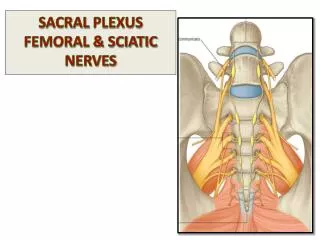
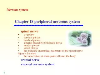
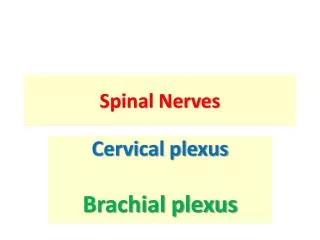
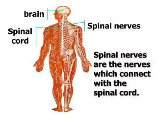
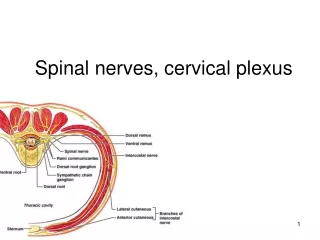
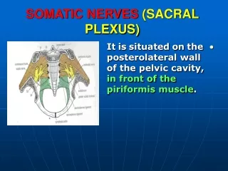
SOMATIC NERVES(SACRAL PLEXUS) • It is situated on the posterolateral wall of the pelvic cavity, in front of the piriformis muscle.
RELATIONS • Anterior : • 1. Parietal pelvic fascia separating it from the internal iliac vessels. • 2. Rectum. • Posterior : • Piriformis muscle.
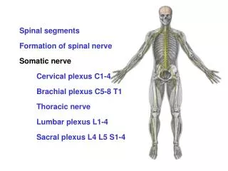
STRUCTURE • It is formed from the ventral rami of (S1- S4) and a contribution from the lumbosacral trunk.
LUMBOSACRAL TRUNK • It is formed of part of the ventral ramus of L4 and all of the ventral ramus of L5. • It descends vertically anterior to the sacroiliac joint.
BRANCHES OF S.P • (A) Passing through the greater sciatic foramen to supply the L.L: • 1. Sciatic nerve (L4-S3). • It is the largest nerve of the plexus and of the body. • It supplies all posterior muscles of back of thigh and leg and all muscles of the sole.
BRANCHES • 2. Superior gluteal nerve (to glutei medius & minimus and tensor fascia lata). • 3. Inferior gluteal nerve (to gluteus maximus). • 4. Nerve to obturatorinternus (and superior gemellus).
BRANCHES • 4. Nerve to quadratus femoris (and inferior gemellus). • 5. Posterior cutaneous nerve of the thigh: • It supplies the skin of the buttock and back of the thigh.
BRANCHES • (B) Branches to the pelvis and perineum:(muscles & viscera) • 1. Pudendal nerve(S2,3 &4) : • It enters the perineum through the lesser sciatic foramen.
BRANCHES • 2. Nerve to piriformis. • 3. Pelvic splanchnic nerves (S2,3 &4). • (c) Perforating cutaneous : to the skin of the lower medial part of the buttock.
OBTURATOR NERVE • It accompanies the lumbosacral trunk to enter the pelvis. • It runs on the lateral pelvic wall.
OBTURATOR NERVE • It lies in the angle between the external and internal iliac vessels. • At the obturator canal, it splits into anterior and posterior divisions that pass through the canal to enter the thigh. • It gives sensory supply to the parietal peritoneum.
AUTONOMIC SUPPLY (PELVIC SYMPATHETIC TRUNK) • It is the continuation of the abdominal trunk. • It is formed of (4- 5) ganglia. • It descends in front of the ala sacrum, medial to the lumbosacral trunk.
(PELVIC SYMPATHETIC TRUNK) • On the anterior surface of the sacrum, it is medial to the • anterior sacral foramina. • It is behind the rectum.
PELVIC SYMPATHETIC TRUNK • Inferiorly, the two trunks unite in front of the coccyx and form a single ganglion (ganglion impar).
BRANCHES • 1. Gray rami communicantes to the lumbar and sacral nerves. • 2. Fibers to hypogastricplexuses.
HYPOGASTRIC PLEXUSES (1) SUPERIOR • Site : in front of the promontry of the sacrum. • Structure : • 1. continuation of the aortic plexuses. • 2. from the 3rd and 4th lumbar ganglia.
SUPERIOR HYPOGASTRIC PLEXUS • Contained fibers: Postganglionic sympathetic, parasympathetic and visceral afferent. • Termination: • It divides into: • Right and Left hypogastric nerves.
(2) INFERIOR HYPOGASTRIC PLEXUS • Site : on each side of rectum, bladder and vagina. • Structure : • Right and left hypogastric nerves. • pelvic splanchnic nerves.
INFERIOR HYPOGASTRIC PLEXUS • Contained fibers : • Postganglionic sympathetic. • Pre and post ganglionic parasympathetic. • Visceral afferent. • Distribution : • To the pelvic viscera through small subsidary plexuses.
PELVIC SPLANCHNIC NERVES • It is the parasympathetic component of the pelvis. • It arises from (S2,3 &4). • The fibers synapse in the inferior hypogastric plexus or in the walls of the viscera. • Some fibers can ascend to the inferior mesenteric plexus and distribute among the branches of the inferior mesentericartery.
EXTERNAL ILIAC ARTERY • Origin : • One of terminal branches of the common iliac artery in front of the sacroiliac joint. • Course : • It descends along the medial border of posoas major. • It enters the thigh by passing deep to the inguinal ligament and becomes the femoral artery.
EXTERNAL ILIAC ARTERY • Branches (in the false pelvis) : • Inferior epigastric. • Deep circumflex iliac.
INTERNAL ILIAC ARTERY • Origin : • The other terminal branch of the common iliac artery. • Course : • It descends inferiorly over the pelvic inlet. • Termination : • At the upper border of the greater sciatic foramen, it divides into anterior and posterior divisions.
POSTERIOR DIVISION • It supplies : • Posterior abdominal wall, posterior pelvic wall and the gluteal region. • Branches : • (1) Iliolumbar artery: • It ascends behind the external iliac vessels, psoas and iliacus. • It divides into lumbar branch (it supplies the muscles of the posterior abdominal wall) and iliac branch for iliacus muscle and bone.
POSTERIOR DIVISION • (2) Lateral sacral arteries : • Usually two. • They descend in front of the sacral plexus. • They give branches that pass into the anterior sacral foramina to supply structures in the vertebral canal, muscles and skin.
POSTERIOR DIVISION • (3) Superior gluteal artery : • It leaves the pelvis through the greater sciatic foramen, above the piriformis. • It supplies the gluteal region.
ANTERIOR DIVISION • It supplies : • Pelvic viscera. • Perineum. • Gluteal region. • Adductor region of the thigh. • The fetus.
ANTERIOR DIVISION • (A) Visceral branches: • 1. Umbilical artery : • It gives the superiorvesicalartery (to the upper part of the urinary bladder) from its patent proximal part. • The distal fibrous part of the artery becomes the medial umbilical ligament.
ANTERIOR DIVISION • 2. Inferior vesical artery : • It supplies the base of the bladder. • In the male it supplies, the prostate and the seminal vesicles. • It gives the artery to the vas deferens.
ANTERIOR DIVISION • (3) Middle rectal artery : • It usually arises with the inferior vesical. • It supplies the muscle layer of the lower rectum . • It anastomoses with the superior and inferior rectal arteries.
ANTERIOR DIVISION • (4) Vaginal artery : • It replaces the inferior vesical artery in the female. • It supplies the base of the bladder and vagina.
ANTERIOR DIVISION • (5) Internal pudendal artery : • It is the main arterial supply to the perineum (skin & muscles) and the anal canal. • It leaves the pelvis through the lower part of the GSF below the piriformis. • It enters the perineum through the LSF.
ANTERIOR DIVISION • (6) Uterine artery : • It runs on the pelvic floor. • It crosses the ureter superiorly. • It passes between the layers of the broad ligament along the lateral margin of the uterus. • It anastomoses with the ovarian artery.
ANTERIOR DIVISION • (B) Parietal branches: • 1. Inferior gluteal : • It leaves the pelvis through the lower part of the GSF below piriformis. • It passes between the sacral nerves (S1- S2) or (S2- S3)
ANTERIOR DIVISION • 2. Obturator artery: • It accompanies the obturator nerve along the lateral pelvic wall. • It leaves the pelvis through the obturator canal.
OVARIAN ARTERY • It arises from the abdominal aorta. • Unlike the testicular artery, it enters the pelvis behind the peritoneum. • It enters the suspensory ligament of the ovary and passes into the broad ligament.
MEDIAN SACRAL ARTERY • It originates from the posterior surface of the aorta just superior to its bifurcation. • It descends in the midline, in front of the anterior surface of the sacrum and coccyx.
EXTERNAL ILIAC VEIN • It begins behind the inguinal ligament as a continuation of the femoral vein. • It ascends along the medial side of its corresponding artery. • It receives the inferior epigastric anddeep circumflex veins.
INTERNAL ILIAC VEIN • It has tributaries corresponding to the branches of the internal iliac artery. • It ascends in front of the sacroiliac joint. • It joins the external iliac vein to form the common iliac vein.
MEDIAN SACRAL VEIN • It accompanies its artery. • It terminates in the left common iliac vein.
LYMPH DRAINAGE • The lymph nodes and vessels are arranged in a chain along the main blood vessels. • They are named according to these vessels: Internal iliac, External iliac and Common iliac.
SACROILIAC JOINTS • They are strong synovial joints. • Articular surfaces : • Auricular surfaces of the sacrum and the iliac bones.
SACROILIAC JOINTS • Stability : • It depends on the strong ligaments : • 1. Posterior. • 2. interosseous sacroiliac ligaments. • The anterior ligament is thin.
SACROILIAC JOINTS • Function : • Transmitting the weight of the body from the vertebral column to the bony pelvis.
ROTATION OF THE SACRUM • The weight of the body tends to thrust the upper end of the sacrum downwards and rotates the lower end upwards.
ROTATION OF THE SACRUM • This is prevented by the following ligaments : • Sacrospinous. • Sacrotuberous. • Iliolumbar : • It is between the tip of the transverse process of L5 and the iliac crest.
SYMPHSIS PUBIS • It is a secondary cartilagenous joint. • The two pubic bones are covered by hyaline cartilage and connected together by a fibrocartilagenous disc. • The joint allows no movement.
SACROCOCCYGEAL JOINT • It is a cartilagenous joint between the bodies of the last sacral vertebrae and the 1st coccygeal vertebra. • It has a great amount of movement.