Download
1 / 25
250 likes | 299 Vues
Explore the complex structure of DNA from nucleotides to base pairing in this informative animation series. Understand the composition, bonding, and functions of the genetic material.

E N D
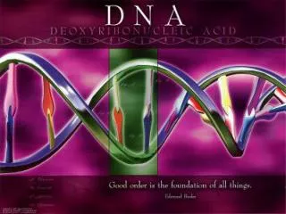
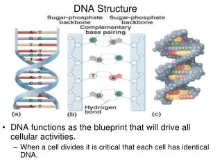
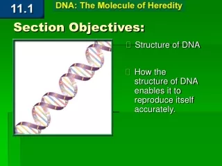
Deepa John Harini Chandra Affiliations Structure of DNA Deoxyribonucleic acid (DNA), which stores and transmits all genetic information, is a long polymer of nucleotide monomers that assumes a complex double helical structure.
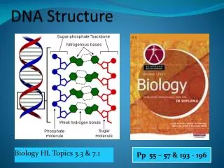
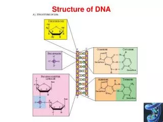
Master Layout (Part 1) 1 This animation consists of 3 parts: Part 1 - Structure of nucleotides: the basic building block of DNA Part 2 - Double helix structure Part 3 - Three forms of DNA Sugar phosphate backbone 2 Thymine Adenine Nitrogenous base Nucleoside 3 Base Base pair Guanine Cytosine Deoxyribose sugar 4 P Base Phosphate Deoxyribose sugar Nucleotide 5 Source: Molecular Biology of the Cell 5/e Garland Science, 2008
Definitions of the components:Part 1 – Structure of nucleotides 1 1. Deoxyribonucleic acid (DNA): A polymer composed of deoxyribonucleotides, linked together by phosphodiester bonds. 2. Nucleotide: The subunit or chain link in DNA or RNA is composed of a sugar, a base and at least one phosphate group. 3. Nucleoside:A base bound to a sugar, either ribose or deoxyribose, by means of a b-glycosidic linkage. 4. Pyrimidine: An organic compound similar to benzene and pyridine that is composed of a heterocyclic, aromatic six-member ring having nitrogen atoms at positions 1 and 3. The nitrogenous bases found in DNA, cytosine and thymine, are derivatives of pyrimidine. 5. Purine: These are the most abundant nitrogen-containing heterocyclic compounds in nature and are composed of a pyrimidine ring fused with an imidazole ring. Derivatives of this aromatic compound occur in DNA in the form of adenine and guanine. 6. Adenine (A): This is a purine base found in both DNA and RNA that pairs with thymine (T) through two hydrogen bonds in the double stranded DNA (dsDNA) structure. In addition to being a component of genetic material, it is also essential for synthesis of various cofactors in the body. 2 3 4 5
Definitions of the components:Part 1 – Structure of nucleotides 1 7. Guanine (G):This is another purine base found in both DNA and RNA. This planar, bicyclic molecule base pairs with cytosine in dsDNA. 8. Cytosine (C): This pyrimidine derivative found to be a component of both DNA and RNA base pairs with Guanine in the dsDNA structure. 9. Thymine (T): Another pyrimidine base derivative that is a component of only DNA where it base pairs with the purine base, adenine. 2 3 4 5
Part 1,Step 1: 1 N-glycosidic linkage Phosphodiester bond Polynucleotide chain 2 3.4 Ao Base Nucleoside Nucleotide 3 3’ 5’ P P P P P Deoxyribose sugar Base Base Base Base 5’ 5’ 5’ 5’ 3’ 3’ 3’ 3’ 4 Action Description of the action Audio Narration (Please use black background) First show the pink pentagon below with label, followed by blue ‘base’ being attached .The curly bracket must appear with the label ‘nucleoside’. Then the ‘pink circle as depicted in the animation must appear followed by the next curly bracket and label ‘nucleotide’. The thick black line must then appear with label followed immediately by the next unit and so on. As shown in animation. DNA is made up of three basic components – a sugar, a nitrogenous base and a phosphate group. The sugar and base are linked to form a nucleoside and attachment of the phosphate group results in a nucleotide. Many such nucleotide units are linked together by means of a covalent bond known as the phosphodiester bond. This is formed between the 3’ carbon of one sugar and 5' carbon of the next sugar via a phosphate group to give rise to a polynucleotide chain. 5
Part 1, Step 2: 1 Guanine Adenine Purines 2 Deoxyribose Deoxyribose Deoxyguanosine Deoxyadenosine 3 Thymine Cytosine Pyrimidines Deoxyribose Deoxyribose 4 Thymidine Deoxycytidine Action Description of the action Audio Narration Show the structures above with their labels and numbering as depicted. (Please use black background & redraw all figures.) Make the four structures appear one at a time as depicted with their labels and numbering around the structure. DNA is composed of four different nitrogenous bases that are derivatives of the heterocyclic, aromatic compounds, purines and pyrimidines. Adenine and guanine are purines while thymine and cytosine are the pyrimidines. The nucleosides of these bases are known as deoxyadenosine, deoxyguanosine, thymidine and deoxyctidine respectively. 5 Source: www.mun.ca/biochem/courses/3107/.../bases_and_chains.html
Master Layout (Part 2) 1 This animation consists of 3 parts: Part 1 - Structure of nucleotides: the basic building block of DNA Part 2 - Double helix structure Part 3 - Three forms of DNA Base pair 2 Minor groove 3 James Watson & Francis Crick Relative absorbance (260nm) Major groove 4 Hydrogen bonding Melting temperature, (Tm) Temperature (oC) 5 Source: www.ncbi.nlm.nih.gov; Biochemistry by Lubert Stryer, 6th edition.
Definitions of the components:Part 2 – Double helix structure 1 1. Base pair:Pairing of complementary bases i.e. A with T and G with C, occurring opposite each other on the two strands of DNA results in formation of a base pair. 2. Hydrogen bonding: These are attractive interactions between hydrogen and a highly electronegative atom like oxygen, nitrogen or fluorine. The hydrogen atom must be flanked by two such electronegative atoms, with one of them involved in covalent bond formation with it. The bases on the opposite strands of DNA are linked together by these relatively weak hydrogen bonds, with A and T forming two hydrogen bonds and G and C forming three. 3. Major and minor groove: The two sugar phosphate backbones of the DNA double helix are not equally spaced along the helical axis. This results in formation of grooves of unequal sizes between the backbone. The wider of the two grooves is known as the major groove while the narrower one is called the minor groove. 4. JamesWatson & Francis Crick: These molecular biologists have been the pioneers in elucidation of the double helical structure of DNA and were awarded the Nobel Prize in 1962 along with Maurice Wilkins. For this reason, the base pairing in DNA is often referred to as “Watson-Crick pairs”. 5. Melting temperature, Tm: The temperature at which half of the DNA strands are in the double helical state while the remaining half are in random coil configuration. This temperature is dependent on both the nucleotide composition of the molecule as well as the length of the molecule. 6. Relative absorbance: The DNA bases absorb UV light strongly at 260 nm and this can be used as a means to detect the amount of DNA. 2 3 4 5
Part 2, Step 1 1 Anti-parallel strands 2 T A Hydrogen bonded base pairs 3 G C DNA double helix Double stranded DNA 4 Audio Narration Action Description of the action (Please use black background & redraw all figures.) First show the left half of the figure on the left appearing with the downward arrow. Next show the right half with the upward arrow coming together with the left half and then the appearance of all the labels. The blue and yellow squares marked ‘A-T’ must be zoomed into and rectangle on top must be shown with appearance of the dotted lines as shown followed by the rectangle at the bottom in the zoomed in mode. It must then be zoomed out and the two strands must then twist around each other to give the figure on the right. Hydrogen bonding between the complementary bases of the two strands of DNA holds them together, with A and T being held together by 2 hydrogen bonds and G and C by 3 bonds. The strands are oriented anti-parallel to each other and twist around an imaginary axis to form the double helical structure. As shown in animation. 5 Source: Molecular Biology of the Cell 5/e Garland Science, 2008; Biochemistry by Stryer, 6th edition
Part 2, Step 2 1 2 Double stranded DNA 3 Double helix unwinding Relative absorbance (260nm) Random coil configuration Hot plate 4 Audio Narration Action Description of the action Melting temperature, (Tm) The process by which two DNA strands of a double helix separate from one another by means of breaking of hydrogen bonds is known as DNA melting or denaturation. Heating of DNA solution causes the strands to separate and the temperature at which half of the DNA strands are in the double helical state while the remaining half are in random coil configuration is known as the melting temperature. The length of the nucleotide sequence & composition of DNA determines the Tm. (Please use black background & redraw all figures.) First show the grey oval with the beaker on top & the solution containing the green & red strands. Next, show the grey oval changing colour to red with a red glow to indicated heating process. While this is happening, the figure must be zoomed into and the green & red strands must be shown to gradually unwind & separate from each other as displayed in the inset. Simultaneously, the graph must also gradually appear on the right with the curve sloping upwards as the strands unwind & once they get separated, the curve must flatten out on the top as shown in the graph. As shown in animation. 5 Temperature (oC) Source: Biochemistry by Stryer, 6th edition
Part 2, Step 3 1 Helical axis 2 Space filling model of DNA double helix Minor groove Major groove 3 4 Action Description of the action Audio Narration The two sugar phosphate backbones of the DNA double helix are not equally spaced along the helical axis. This results in formation of grooves of unequal sizes between the backbone. The wider of the two grooves is known as the major groove while the narrower one is called the minor groove. Zoom into the depicted portion of the structure above and show the figure on the right with its labels clearly indicated. (Please use black background & redraw all figures.) First show the structure above rotating about the axis in the direction indicated. Then zoom into the depicted region and show the labels. 5 Source: Molecular Biology of the Cell 5/e Garland Science, 2008
Part 2, Step 4: 1 Chargaff’s Rule: C T G A A G C T G A A G T C T C A G T C 2 3 % Adenine = % Thymine % Guanine = % Cytosine 4 Action Description of the action Audio Narration (Please use black background) First show the weighing balance on the left appearing followed by the pink and blue figures on top. First the pink must enter the weighing pan and the balance must tilt to the left. Then the blue must enter the right pan and the balance must get stabilized again. Similarly for the figure on the right. Chargaff’s rule states that DNA from any organism must have a 1:1 ratio of purine and pyrimidine bases. More specifically, it states that the amount of adenine is always equal to the amount of thymine and amount of guanine is equal to cytosine. As shown in animation. 5
Master Layout (Part 3) 1 This animation consists of 3 parts: Part 1 - Structure of nucleotides: the basic building block of DNA Part 2 - Double helix structure Part 3 - Three forms of DNA 2 3 4 A- DNA Z- DNA B- DNA 5 Source: Genetics by Peter J.Russel
Definitions of the components:Part 3 – Three forms of DNA 1 1. A-DNA:A-DNA is one of the many possible double helical structures that DNA can assume. It is a right-handed double helix that is observed when the humidity is relatively low. 2. B-DNA: The double helix structure deduced by Watson & Crick that is normally observed in most organisms is the B-DNA. This is also a right-handed double helix but is observed when humidity is relatively high. 3. Z-DNA: This is a left handed double helical structure of DNA in which the double helix is found to wind to the left in a zig-zag pattern. 2 3 4 5
Part 2, Step 1 1 Overall helix proportions Elongated and thin Short and wide Longer and thinner Helix rotation sense Helical axis Base pairs/turn of helix 2 Very narrow & deep Very broad, shallow Narrow, medium depth Minor groove 12.0 10.0 3 10.9 Flattened on helix surface Very narrow & deep Wide, medium depth Major groove A- DNA B-DNA Z- DNA 4 Action Description of the action Audio Narration (Please use black background & redraw all figures.) First show the three structures of above with the dotted line depicting the axis. Next, show the parameters on the left (in bold) appearing one at a time along with their respective description for each structure as depicted in the animation. DNA exists in many possible conformations that include A-DNA, B-DNA, and Z-DNA forms. A and B forms are right handed helices whereas Z- DNA is a left handed helix. There are 10.9,10.0 and 12.0 base pairs per helix turn in A,B and Z-DNA forms respectively. They differ in their overall structural proportions as well in the proportions of their major and minor grooves. As shown in animation. 5 Source: Genetics by Peter J.Russel
Interactivity option 1:Step No: 1 1 Shown below is a single strand of DNA which needs to base paired with its complementary strand. The four bases of DNA are shown at the bottom. Select the correct complementary base for each of the position of this strand. 2 C T A T G A A C C G G T A C T 3 A G G C T A C T T G A C A T G Adenine Cytosine C G Thymine Guanine T A 4 Results Interacativity Type Boundary/limits Options Once the user matches all the As with Ts and Gs with Cs, the two strands must come together and the strand must fit into the grooves on the bottom strand. Correct sequence which the user must get is displayed above. If any mistake is made, it must be circled in red. The strand at the bottom must be shown and the four shapes on the right must be shown. User needs to drag and drop these shapes into their corresponding positions on the the strand shown on top. Drag and drop. 5
Interactivity option 2:Step No:1 1 • Average length of double helix in a human chromosome is 3.8cm.What is its molecular weight? 2 A) 1.01×10⁸ daltons B) 7.37×10¹⁰ daltons C) 10.1×10⁸ daltons 3 D)73.7×10¹⁰ daltons Interacativity Type Options Boundary/limits Results User has to choose one of the four Options. If A or C or D chosen, then they must turn red. User can however continue till he gets the right answer(B), which must turn green. User is then directed to step 2. B is the right answer. User has to choose one of the four Options. If A, C or D are chosen, then they must turn red. User can however continue till he gets the right answer(B) which must turn green. User is then directed to step 2. Choose the correct option 4 5
Interactivity option 2:Step No:2 One base pair occupies 0.34nm or 0.34 × 107 cm in the double helix, so the number of base pairs in the chromosome is 3.8 divided by 0.34 × 10⁷ or 1.117 × 10⁸ base pairs. Each base pair on an average, weighs 660 daltons, and therefore the molecular weight of chromosome is : 1.117 × 108 × 660 = 7.37×10¹⁰ daltons
Interactivity option 3:Step No: 1 1 Three classical experiments, by Frederick Griffith, Avery-MacLeod-McCarty and Hershey & Chase, carried out soon after the discovery of DNA are described below. What inference can be made from these experiments? a) That proteins are the transforming elements responsible for hereditary characteristics 2 b) That RNA is the transforming element responsible for hereditary characteristics 3 c) That DNA is the transforming element responsible for hereditary characteristics d) That proteins and RNA act together as the transforming element 4 Results Interacativity Type Boundary/limits Options Option ‘c’ is the correct answer. If the user chooses option ‘c’, message saying ‘correct answer’ must be displayed. If user chooses any of the other options, message saying ‘incorrect answer’ must be displayed. (Please redraw all figures) User must be allowed to choose one of the options given above after he views the animations described in the subsequent slides. The options must be displayed only on completion of the animations. Choose the correct option. 5
Interactivity option 3:Step No: 2(a) 1 Experiment by Hershey & Chase T2 phage particle Radioactive 35S labeled protein coat Non-radioactive DNA 2 Infection 3 E. coli cell E. coli cell T2 phage particle Radioactive 32P labeled DNA Non-radioactive protein coat 4 Infection 5 E. coli cell E. coli cell
Interactivity option 3:Step No: 2(b) 1 Experiment by Hershey & Chase Treatment in blender to remove protein coat No radioactivity in bacterial cell pellet Radioactivity in supernatant 2 Centrifugation Non-radioactive progeny phage 3 Infected E. coli cell Treatment in blender to remove protein coat Radioactivity in bacterial cell pellet No radioactivity in supernatant 4 Centrifugation Radioactive progeny phage 5 Infected E. coli cell
Interactivity option 3:Step No:2 (c) Experiment by Fredrick Griffith, Avery-MacLeod-McCarty Inject mice Dies, Type III S virulent bacteria recovered Type III S Streptococcus pneumoniae: Living virulent Inject mice Survives, no bacteria recovered Type II R: Living non-virulent Inject mice Survives, no bacteria recovered Type III S: Heat killed Dies, Type III S virulent bacteria recovered Inject mice Type II R: Living, non-virulent Type III S: Heat killed
Interactivity option 3:Step No:2(d) Add DNA to R bacteria Treat with RNase Plate on growth medium S transformants produced Mixture of DNA and RNA Add RNA to R bacteria Plate on growth medium Treat with DNase No S transformants produced Mixture of DNA and RNA
Questionnaire 1 1. Nucleotides contain _____. Answers: a) sulphur b) nitrogen base c) a 6- carbon sugar d)All the above 2. Avery's research showed that DNA is the transforming substance based on the observation that _____ Answers: a) enzymes that degrade proteins did not prevent transformation b) RNase, an enzyme that digests RNA, did not prevent transformation c) Dnase, an enzyme that digests DNA did prevent transformation d)all of the above 3.The structure of DNA was described _____. Answers: a) in the 150’s b) in 1900 c) in 1950’s d) in 1990 4.Which of these blocks phosphodiester bonding thereby blocking chain elongation? Answers: a) Rifamycin b) Rifampicin c) Alpha amanitin d) Chloramphenicol 5. Z-DNA Answers: a) Winds to the left b) Is thicker than B-DNA c) Winds to the right d)Has a broad minor groove 2 3 4 5
Links for further reading Books: • Genetics by Peter J. Russell, 5th edition Research papers: • A Structure for Deoxyribose Nucleic AcidWatson J.D. and Crick F.H.C. Nature 171, 737-738 (1953)