Download
1 / 46
470 likes | 648 Vues
Acquired alterations of IGH and TCR loci in lymphoproliferative disorders. Sabine Franke, PhD CHU Liège, Centre for Human Genetics. Interuniversity Course - HUMAN GENETCIS. 23/04/10 ULG. Overview. what are lymphoproliferative disorders?

E N D
Acquired alterations of IGH and TCR loci in lymphoproliferative disorders Sabine Franke, PhD CHU Liège, Centre for Human Genetics Interuniversity Course - HUMAN GENETCIS 23/04/10 ULG
Overview • what are lymphoproliferative disorders? • what is Immunoglobulin (IG) and T-cell receptor (TCR)? • what are alterations and how to detect them? • examples of clinical cases
Blood cell development 2 major types of lymphocytes: B and T cells
Lymphoproliferative disorders (LPDs)LPDs refer to several conditions in which lymphocytes are produced in excessive quantities. • Chronic lymphocytic leukemia • Acute lymphoblastic leukemia • Hairy cell leukemia • lymphomas • Multiple myeloma • Waldenstrom’s macroglobulinemia • Wiskott-Aldrich syndrome • Post-transplant lymphoproliferative disorder • Autoimmune lymphoproliferative syndrome (ALPS) • ‘Lymphoid interstitial pneumonia’
B, T, and NK lineage of lymphoid malignancies Lymphoid malignancy B lineage T lineage Natural Killer lineage Acute lymphoblastic leukemia– children 82 – 86% 14 – 18% < 1%– adults 75 – 80% 20 – 25% < 1% Chronic lymphocytic leukemias 95 – 97% 3 – 5% 1 – 2% Non-Hodgkin lymphomas – nodal NHL 95 – 97% 3 – 5% < 2%– extranodal NHL 90 – 95% 5 – 10% < 2%– cutaneous NHL 30 – 40% 60 – 70% < 2% Multiple myeloma 100% 0% 0%
Overview • what are lymphoproliferative disorders? • what is Immunoglobulin (IG) and T-cell receptor (TCR)? • what are alterations and how to detect them? • examples of clinical cases
Schematic representation of an immunoglobulin
Gene rearragements of the antigen receptor genes occur during the lymphoid proliferation
IG segments Leukemic B-cells have a specific V-D-J segment encoding the Ig protein. PCR can be used to take advantage of this trait to detect leukemia clones. Use "forward" and "reverse" primers that flank the region.
! • These gene rearrangements generate products that are unique in length and sequence in each cell.
Estimated diversity of human Ig and TCR molecules IgH Igα Igג TCR αβγδ molecules molecules Number gene segments V gene segments ~44 ~43 ~38 ~46 ~47 ~6 ~6 D gene segments 27 2 3 J gene segments 6 5 4 53 13 5 4 Combination diversity >2x10 6 2x10 6 <5000 Junctional diversity ++ ++ + ++ ++ ++++ Total diversity >10 12 >10 12 >10 12
The molecular synthesis of the Immunoglobulins (Ig) and T cell Receptors (TcR) includes complex mechanisms such as DNA rearrangements, nucleotide insertions and deletions, and for the Ig, somatic hypermutations. This leads one individual to potentially produce more than 1012 different Ig or TcR.
Overview • what are lymphoproliferative disorders? • what is Immunoglobulin (IG) and T-cell receptor (TCR)? • what are alterations and how to detect them? • examples of clinical cases
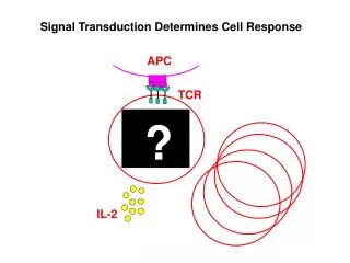
! Identify lymphocyte populations derived from one single cell using the unique V-J gene rearrangements present within these antigen receptor loci.
Variation of IGH genes stepwise rearrangement of V, D, and J gene segments The genes encoding antigen receptors are unique because of their high diversity and their assembly in developing lymphocytes from gene segments through a series of site-specific DNA recombination reactions known as V(D)J rearrangement. (junctional region) ok
T-cell receptor gene rearrangement The variable domain of both the TCR α-chain and β-chain have three hypervariable or complementary determining regions (CDRs) • Receptor for antigen on the majority of mature T-cells consists of two polypeptides alpha and beta that are linked by disulphide bonds and are associated with CD3 • A small population of mature T-cells express a different TCR heterodimer in association with CD3. This is composed of two polypeptides designated gamma and delta ok
Clinically relevant testing • Reactive versus malignant • B-cell versus T-cell malignancy • New lymphoma versus recurrence • Assessment of remission and relapse • Clinically relevant: bone marrow involvement • (relation with prognosis) • Evaluation of treatment effectiveness • detection of minimal residual disease - treatment
Analysis methods • PCR (Polymerase chain reaction) - B-cell: Ig rearrangements - T-cell: TCR rearrangements • Sequencing • Southern blot • (FISH: fluorescence in situ hybridization)
PCR : Discrimination between monoclonal and polyclonal Ig/TCR gene PCR products • GeneScanning analysis Fast, accurate, sensitive non-quantitative monitoring of clonal proliferations Need sequence equipment • Heteroduplex analysis Sensitivity ~5-10% available to most laboratories
Design of novel primer sets for detection of Ig/TCR rearrangements BIOMED-2 study (multiplex PCR) Ig genes: IGH: VH-JH and DH-JH IGK: V-J and Kde rearrangements IGL: V-J TCR genes: TCRB: V-J and D-J TCRG: V-J TCRD: V-J, D-D, D-J, and V-D
BIOMED-2 clonality strategy Suspected lymphoma of unknown origin Suspected T-cell lymphoma Suspected B-cell lymphoma TCRGVJ (A and B) TCRB V(D)J (A and B) TCRB DJ IGH V(D)J FR1, FR2, FR3 IGH DJ(A) IGK-VJ and DE
In order to amplify the DNA by PCR successfully, the primers must recognize DNA sequences within a short segment of DNA. In the germline configuration, the V and J segments are widely separated, and the primers will be so far apart that the enzyme cannot synthesize new DNA and no PCR product is seen. In a polyclonal B cell population, you would see a smear, since each B-cell would have a different V-D-J length (and sequence). However, if the B-cells are from one clone, they will all have the same V-D-J size segment and therefore only one band.
PCR GeneScan analysis region region
Example BIOMED-2 multiplex IGHVH-FR2–JH Use of controls
From the patient to the analysis • Selection of material • protocol • Check DNA quality • Clonality analysis
Overview • what are lymphoproliferative disorders? • what is Immunoglobulin (IG) and T-cell receptor (TCR)? • What are alterations and how to detect them? • examples of clinical cases
Examples of clinical cases • Lymphoma? • Relapse?
Case 1 • Female • 63 years • B-cell lymphoma in dec. 2004 • FR1 polyclonal, FR3 monoclonal • Feb. 2009 biopsy • Relapse?
FR3 GeneScan analysis of controls IGH FR1 FR2 FR3 DH JH Control GeneScan analysis H2O GeneScan analysis of sample GeneScan analysis of polyclonal control
Case 1: Relapse? FR3 2/2009 Biopsie FR3: 152,40 bp 12/2004 Biopsie FR3: 152,35 bp 2000 12/2004 600
Results of the 1. case Controls: ok B-cell targets: IGH(VDJ) FR3 monoclonal Molecular conclusion: Clonal rearrangement of the IGH gene was detected in this specimen. This gene rearrangement profile is identical to the one detected in the biopsie of 12/2004 and confirms the relapse of the disease. For confirmation sequencing can be done
Case 2 • Female • 81 years • biopsie • Lymphoma?
Case 2 3/2009 biopsie FR1 327,11 bp 2700 Polyclonal control 100
Results of 2. case Controls: ok B-cell targets: IGH(VDJ) FR1 monoclonal FR2 monoclonal Molecular conclusion: Clonal rearrangements of the IGH gene were detected in this specimen. This gene rearrangement profile fits to the presense of a monoclonal B-cell population/B-NHL.
Use of clonality analysis 1. Making the diagnosis Normal reactive malignant 2. Involvement (staging) 3. Assessment of remission and relapse Normal reactive malignant 4. Evaluation of treatment effectiveness - detection of minimal residual disease (MRD) MRD-based risk-group stratification (treatment reduction or treatment intensification)
Detection of abnormality at molecular level • PCR • Southern blot • Sequencing • FISH
Southern blot <-> PCR • Small region • Less DNA • Very sensitive • Automatisation • Fast • Ethidium bromide? • Large region • Large quantity DNA • Less sensitive • Labor intensive • Takes several days • radioactice
FISH – Fluorescence in situ hybridization • Relative large region • Translocation partner has not te be known • On metaphases and nuclei
FISH • Excellent diagnostic tool for detection of well-defined chromosome aberrations
Summary • Gene rearrangements of the antigen receptor genes occur during the lymphoid proliferation • These gene rearrangements generate products that are unique in length and sequence in each cell. B-cells: Ig T-cells: TCR • Unique length allows by PCR discrimination of monoclonality and polyclonality