Download
1 / 18
180 likes | 226 Vues
Learn about Electron Microscopes, their uses, Crystallography, and how SEM and TEM instruments work. Discover the fascinating world of microscopy.

E N D
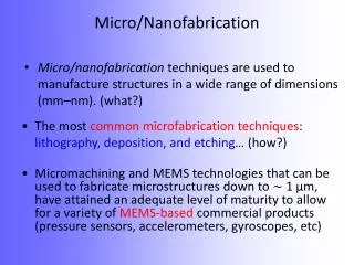
NANO 225 Micro/NanoFabrication Electron Microscopes
Microscopes • Electron Microscopes • Magnification: 1,000,000 X • Resolution: <1 nm • down to 0.5 A (TEM) • Use focused beam of electrons instead of light • “Lenses” are coils, not glass • Sample in vacuum Light Microscopes • Magnification: 500 X to 1000 X • Resolution: 0.20 µm • Limits reached by early 1930s • Color images • Sample in air • Scanning Electron Microscope (SEM) • Transmission Electron Microscope (TEM)
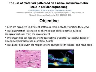
Electron Microscopes What can electron microscopes tell us? • Morphology • Size and shape • Topography • Surface features (roughness, texture, hardness) • Crystallography • Organization of atoms in a lattice
Electron Microscopes: Crystallography Crystallography: • Arrangement of atoms • Crystals have atoms arranged in ordered lattices • Amorphous: no ordering of atoms Crystallography affects properties (electrical, strength, etc)
Scanning Electron Microscopy (SEM) • Provides information about: • Topography of sample or structure • Chemical composition near the surface of sample • Magnification: ~30X to 500,000X • Resolution • Nanometer scale • Dependent on: • wavelength of electrons () • Numerical aperture of lens system (NA) • Electron gathering ability of the objective • Electron providing ability of the condenser
SEM Instrument • Electron beam • Spot size ~5 nm • Energy ~200 - 50,000 eV (electron volts) • Rastered over surface of sample • Emitted electrons collected on a cathode ray tube (CRT) to produce SEM images • Sample Prep: • Attach to Al “stub” with conductive carbon tape or paste • Sputter-coat non-conductive samples
SEM: How it works • Electron beam strikes surface and electrons penetrate surface • Interactions occur between electrons and sample • Electrons and photons emitted from sample • Emitted electrons captured on CRT • SEM image made from detected electrons http://www.youtube.com/watch?v=bfSp8r-YRw0&feature=related http://www.youtube.com/watch?v=fToTFjwUc5M&feature=related
SEM: Electron Beam Interactions Valence electrons • Inelastic scattering: Energy transferred to atomic electron • If atomic electron has high enough energy can be emitted from sample • “Secondary electron” if energy of emitted electron <50 eV Atomic nuclei • “Backscattered electrons” • Elastic scattering: e- bounce off with same amount of energy • Atoms with high atomic numbers cause more backscattering Core electrons • Core electron ejected from sample; atom becomes excited • To return to ground state, x-ray photon or Auger electron emitted
Transmission Electron Microscopy (TEM) • Provides information about: • Topography of sample or structure • Chemical composition • Magnification: ~50X to 1,000,000X • Resolution • Dependent on: • Electron mass (m) and charge (q) • Potential difference used to accelerate electrons (V) • Proportional to 3/4 • < 0.2 nm resolution (400 keV)
TEM Instrument • Electron beam • Energy: 100,000 - 1,000,000 eV (100 keV - 1 MeV) • Projected onto thin sample using lens system by deflection coils • Sample Prep: • Need very thin sample! • Slice of bulk material or cross-section of thin film • Grind into power, dissolve, put on conductive grid to evaporate http://www.vcbio.science.ru.nl/images/10-tem_grid_zoom.jpg http://video.google.com/videoplay?docid=-5489601762301542658&q=transmission+electron+ microscope&total=6&start=0&num=10&so=0&type=search&plindex=1
TEM: Electron Beam Interactions Electron beam strikes surface and is transmitted through film Scattering occurs during transmission Unscattered electrons pass through the sample and are detected (along with elastic scattered electrons) Elastic Scattering • No energy loss • Diffraction patterns Inelastic Scattering • Occurs at heterogeneties (defects, grain boundaries)
SEM and TEM Instruments http://www.vcbio.science.ru.nl/en/image-gallery/electron/
SEM and TEM Comparison • SEM makes clearer images than TEM • SEM has easier sample preparation than TEM • TEM has greater magnification than SEM • SEM has large depth of field
SEM and TEM Data Images • Ag thin film deposited on Si substrate (thermal or e-beam evaporation) • TCNQ (7,7,8,8-tetracyanoquinodimethane) powder and Ag thin film are enclosed in a vacuum glass tube, then heated in a furnace. http://nami.eng.uci.edu/projects/Agtcnq.htm
Some definitions • Stigmation: • correcting asymmetries in horizontal v. vertical focus • seen as “streakiness” • Collimation: • creation of parallel path particles • typically no control over
Improving Images: Spot size Spot size: electron spot radius (rms) • Especially useful to improve focus at high mag • Minimize spot size: • Decrease working distance • Increase current on focusing lens Trade-offs: • Smaller area covered • Lower beam current (worse contrast)
Improving Images: Depth of field Depth of field: • How many planes are in focus at once • Related to distance that beam stays narrow • Especially useful to see detail on rough surfaces: • Maximize DOF: • Decrease aperture size • Decrease magnification • Increase working distance Trade-off: • Lower magnification
Improving Images: Signal-to-Noise Signal-to-noise ratio: contrast between interacting and non-interacting surfaces • Especially useful to gain more fine detail • Maximize S/N ratio: • High beam current • Slow scan rate Trade-off: • Much larger spot size