Pedigree Charts
Pedigree Charts. The family tree of genetics. What is a Pedigree?. A pedigree is a chart of the genetic history of family over several generations. Scientists or a genetic counselor would find out about your family history and make this chart to analyze. Male. Female.

Pedigree Charts
E N D
Presentation Transcript
Pedigree Charts The family tree of genetics
What is a Pedigree? • A pedigree is a chart of the genetic history of family over several generations. • Scientists or a genetic counselor would find out about your family history and make this chart to analyze.
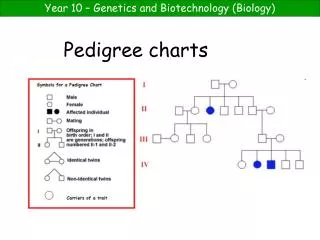
Male Female Constructing a Pedigree
Connecting Pedigree Symbols Examples of connected symbols: • Married Couple • Siblings
Connecting Pedigree Symbols Examples of connected symbols: • Fraternal twins • Identical twins
Marriage Has the trait Female-daughter Female-daughter Male-Son Male- Son Oldest to youngest Male-DAD Female-MOM
Interpreting a Pedigree Chart • Determine if the pedigree chart shows an autosomal or X-linked disease. • If most of the males in the pedigree are affected the disorder is X-linked • If it is a 50/50 ratio between men and women the disorder is autosomal.
Example of Pedigree Charts • Is it Autosomal or X-linked?
Answer • Autosomal
Interpreting a Pedigree Chart • Determine whether the disorder is dominant or recessive. • If the disorder is dominant, one of the parents must have the disorder. • If the disorder is recessive, neither parent has to have the disorder because they can be heterozygous.
Example of Pedigree Charts • Dominant or Recessive?
Answer • Dominant
Example of Pedigree Charts • Dominant or Recessive?
Answer • Recessive
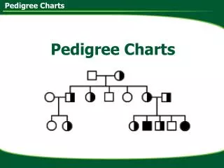
ff ff Ff ff Ff Key: affected male affected female unaffected male unaffected female Ff • Steps: • Identify all people who have the trait. • For the purpose of this class all traits will be given to you. In other instances, you would have to determine whether or not the trait is autosomal dominant, autosomal recessive, or sex-linked. • In this example, all those who have the trait are homozygous recessive. • Can you correctly identify all genotypes of this family? • F- Normal • f- cystic fibrosis
Key: affected male affected female unaffected male unaffected female Pp Pp • PKU • P- Unaffected • p- phenylketonuria Pp pp PP or Pp pp pp Pp Pp
Key: affected male affected female unaffected male unaffected female hh Hh • H-huntington’s disease • h-Unaffected Hh hh Hh hh hh Hh hh
Summary • Pedigrees are family trees that explain your genetic history. • Pedigrees are used to find out the probability of a child having a disorder in a particular family. • To begin to interpret a pedigree, determine if the disease or condition is autosomal or X-linked and dominant or recessive.
Autosomal Dominant Polydactyly HD Achondroplasia Progeria
Autosomal Recessive • Disorders • Cystic Fibrosis • Tay-sachs Disease
Karyotypes • To analyze chromosomes, cell biologists photograph cells in mitosis, when the chromosomes are fully condensed and easy to see (usually in metaphase). • A picture of chromosomes arranged in this way is known as a karyotype.
Karyotypes • The karyotype is a result of a haploid sperm (23 chromosomes) fertilizing a haploid egg (23 chromosomes). • The diploid zygote (fertilized egg) contains the full 46 chromosomes. (in humans)
Labeling a Karyotype • To label a karyotype correctly, first list the number of chromosomes found in the karyotype. Ex. 46 • Secondly, list the type of sex chromosomes found in the karyotype. Ex. XX • Lastly, list the any abnormalities at the appropriate chromosome number. Normal Human Female: 46, XX Normal Human Male: 46, XY
What are abnormalities? • Sometimes, during meiosis, things go wrong. • The most common error is nondisjunction, which means “not coming apart”. • If nondisjunction occurs , abnormal numbers of chromosomes may find their way into gametes, and a disorder of chromosome numbers may result.
Autosomal Chromosome Disorders • Two copies of an autosomal chromosome fail to separate during meiosis, an individual may be born with THREE copies of a chromosome. • This is known as a “Trisomy” • Trisomy 13, Trisomy 18, Trisomy 21.
Male: 47, XY, +21 Female: 47, XX, +21 Down Syndrome • Most common, Trisomy 21 (down syndrome) • 1 in 800 babies born in U.S. with Trisomy 21. • Mild to severe mental retardation • Increased susceptibility to many diseases and a higher frequency of other birth defects.
Klinefelter’s Syndrome, 47 XXY Sex Chromosome Disorders • Turner’s Syndrome (nondisjunction) • Female inherits only one X chromosome • Karyotype: 45, X • Women are sterile, sex organs do not develop at puberty. • Klinefelter’s syndrome (nondisjunction) • Males receive an extra X chromosome • Karyotype: 47, XXY • The extra X chromosome interferes with meiosis and prevents ind. from reproducing.