Download
1 / 49
580 likes | 1.56k Vues
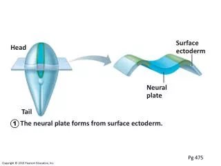
Surface ectoderm . Head. Neural plate . Tail. The neural plate forms from surface ectoderm. 1. Pg 475. Neural folds. Neural groove . The neural plate invaginates, forming the neural groove, flanked by neural folds. 2. Pg 475. Neural crest.

E N D
Surface ectoderm Head Neural plate Tail The neural plate forms from surface ectoderm. 1 Pg 475
Neural folds Neural groove The neural plate invaginates, forming the neural groove, flanked by neural folds. 2 Pg 475
Neural crest Neural fold cells migrate to form the neural crest, which will form much of the PNS and many other structures. 3 Pg 475
Head Surface ectoderm Neural tube Tail The neural groove becomes the neural tube, which will form CNS structures. 4 Pg 475
(a) Neural tube (e) Adultneuralcanalregions (b) Primary brainvesicles (c) Secondary brainvesicles (d) Adult brainstructures Neocortex Cerebrum: cerebral hemispheres (cortex, white matter, basal nuclei) Lateral ventricles Telencephalon Mammalian Diencephalon (thalamus, hypothalamus, epithalamus), retina Anterior (rostral) Prosencephalon (forebrain) Third ventricle Diencephalon Emotion Mesencephalon (midbrain) Cerebral aqueduct Brain stem: midbrain Mesencephalon Rhombencephalon (hindbrain) Metencephalon Brain stem: pons Fourth ventricle Cerebellum Brain stem: medulla oblongata Myelencephalon Primitive brain Reflexes Posterior (caudal) Central canal Spinal cord Pg 429
Anterior (rostral) Posterior (caudal) Metencephalon Mesencephalon Midbrain Flexures Diencephalon Cervical Telencephalon Spinal cord Myelencephalon (a) Week 5 Pg 430
Cerebral hemisphere Outline of diencephalon Midbrain Cerebellum Pons Medulla oblongata Spinal cord (b) Week 13 Pg 430
Cerebral hemisphere Cerebellum Pons Medulla oblongata Spinal cord (c) Week 26 Pg 430
Cerebral hemisphere Diencephalon Cerebellum Brain stem • Midbrain • Pons • Medullaoblongata (d) Birth 3 Lbs. Gelatinous mass Pg 430
Anterior Longitudinal fissure Frontal lobe Cerebral veins and arteries covered by arachnoid mater Parietal lobe Right cerebral hemisphere Left cerebral hemisphere Occipital lobe Posterior (c) Cerebrum Pg 432
. Commissural fibers (corpus callosum) Longitudinal fissure Superior Lateral ventricle Association fibers Basal nuclei • Caudate Corona radiata • Putamen • Globuspallidus 2 mm True dendrites and cell bodies Convoluted Fornix Internal capsule Thalamus Gray matter/Cortex Third ventricle White matter Projection fibers Pons Decussation of pyramids Medulla oblongata (a) Pg 438
Precentral gyrus Central sulcus Postcentral gyrus Frontal lobe Parietal lobe Parieto-occipital sulcus (on medial surface of hemisphere) Lateral sulcus Occipital lobe Temporal lobe Transverse cerebral fissure Cerebellum Pons Medulla oblongata Fissure Spinal cord (a deep sulcus) Gyrus Cortex (gray matter) Sulcus-Shallow groove White matter (a) Pg 432
Motor areas Sensory areas and related association areas Central sulcus 2 Primary motor cortex 10 Primary somatosensory cortex 3 Premotor cortex Somatic sensation 4 Frontal eye field Somatosensory association cortex Broca’s area (outlined by dashes) 5 11 Gustatory cortex (in insula) Taste 1 Prefrontal cortex Wernicke’s area (outlined by dashes) Working memory for spatial tasks 8 Executive area for task management Primary visual cortex 12 Working memory for object-recall tasks Vision Visual association area Solving complex, multitask problems 13 Auditory association area 7 Hearing Primary auditory cortex 6 (a) Lateral view, left cerebral hemisphere Motor association cortex Primary sensory cortex Primary motor cortex Sensory association cortex Multimodal association cortex Angular Gyrus Gnostic Area -sensory to prefrontal cortex Pg 434
. Posterior -Voluntary -Skeletal Muscle -Spacial Motor Sensory Anterior Motor map in precentral gyrus Sensory map in postcentral gyrus Discrete Motor Control Toes Genitals Jaw Primary motor cortex (precentral gyrus) Primary somato- sensory cortex (postcentral gyrus) Tongue Intra- abdominal Swallowing -Facilitory Impulses -Contralateral -Damage 435
Motor areas Sensory areas and related association areas Central sulcus 2 Primary motor cortex 10 Primary somatosensory cortex 3 Premotor cortex Somatic sensation 4 Frontal eye field Somatosensory association cortex L Broca’s area (outlined by dashes) 5 Motor aphasia 11 Gustatory cortex (in insula) Taste 1 Prefrontal cortex Wernicke’s area (outlined by dashes) Working memory for spatial tasks 8 Executive area for task management Primary visual cortex 12 Working memory for object-recall tasks Vision Visual association area Solving complex, multitask problems 13 Auditory association area 7 Hearing Primary auditory cortex 6 (a) Lateral view, left cerebral hemisphere Motor association cortex Primary sensory cortex Primary motor cortex Sensory association cortex Multimodal association cortex Angular Gyrus Gnostic Area -sensory to prefrontal cortex Pg 434
Motor areas Sensory areas and related association areas Central sulcus 2 Primary motor cortex 10 Primary somatosensory cortex 3 Premotor cortex Somatic sensation 4 Frontal eye field Somatosensory association cortex Broca’s area (outlined by dashes) 5 11 Gustatory cortex (in insula) Taste 1 Prefrontal cortex Wernicke’s area (outlined by dashes) Working memory for spatial tasks 8 Executive area for task management Primary visual cortex 12 Working memory for object-recall tasks Vision Visual association area Solving complex, multitask problems 13 Auditory association area 7 Hearing Primary auditory cortex 6 (a) Lateral view, left cerebral hemisphere Motor association cortex Primary sensory cortex Primary motor cortex Sensory association cortex Multimodal association cortex Angular Gyrus Gnostic Area -sensory to prefrontal cortex Pg 434
-Hear what you see -See what you hear Primary Motor Cortex Puts words in order Fluent Aphasia (Word Salad) Agraphia Alexia
Cingulate gyrus Primary motor cortex Premotor cortex Central sulcus Corpus callosum Primary somatosensory cortex Frontal eye field Parietal lobe Somatosensory association cortex Prefrontal cortex Parieto-occipital sulcus Occipital lobe Processes emotions related to personal and social interactions Visual association area Orbitofrontal cortex Olfactory bulb Primary visual cortex Olfactory tract Fornix Uncus Calcarine sulcus Temporal lobe Primary olfactory cortex Parahippocampal gyrus (b) Parasagittal view, right hemisphere Motor association cortex Primary sensory cortex Primary motor cortex Sensory association cortex Multimodal association cortex Pg 434
Motor areas Sensory areas and related association areas Central sulcus 2 Primary motor cortex 10 Primary somatosensory cortex 3 Premotor cortex Somatic sensation 4 Frontal eye field Somatosensory association cortex Broca’s area (outlined by dashes) 5 11 Gustatory cortex (in insula) Taste 1 Prefrontal cortex Wernicke’s area (outlined by dashes) Working memory for spatial tasks 8 Executive area for task management Primary visual cortex 12 Working memory for object-recall tasks Vision Visual association area Solving complex, multitask problems 13 Auditory association area 7 Hearing Primary auditory cortex 6 (a) Lateral view, left cerebral hemisphere Motor association cortex Primary sensory cortex Primary motor cortex Sensory association cortex Multimodal association cortex Angular Gyrus Gnostic Area -sensory to prefrontal cortex Pg 434
Posterior -contralateral -Locates -Determines kind and strength of sensation -Projects -Damage Motor Sensory Anterior Motor map in precentral gyrus Sensory map in postcentral gyrus Toes Genitals Jaw Primary motor cortex (precentral gyrus) Primary somato- sensory cortex (postcentral gyrus) Tongue Intra- abdominal Swallowing Pg 435
Motor areas Sensory areas and related association areas Central sulcus 2 Primary motor cortex 10 Primary somatosensory cortex 3 Premotor cortex Somatic sensation 4 Frontal eye field Somatosensory association cortex Broca’s area (outlined by dashes) 5 11 Gustatory cortex (in insula) Taste 1 Prefrontal cortex Wernicke’s area (outlined by dashes) Working memory for spatial tasks 8 Executive area for task management Primary visual cortex 12 damage Working memory for object-recall tasks Vision Visual association area Solving complex, multitask problems 13 Auditory association area 7 Hearing Primary auditory cortex 6 (a) Lateral view, left cerebral hemisphere Motor association cortex Primary sensory cortex Primary motor cortex Sensory association cortex Multimodal association cortex Angular Gyrus Gnostic Area -sensory to prefrontal cortex Pg 434
Commissural fibers (corpus callosum) Longitudinal fissure Superior Lateral ventricle Association fibers Basal nuclei (Ganglia) • Caudate Corona radiata • Putamen • Globuspallidus Fornix Internal capsule Thalamus Gray matter Third ventricle White matter Projection fibers Pons -Medulla -White Matter -Myelinated axons -Oligodendrocytes Decussation of pyramids Medulla oblongata (a) Pg 438
Fibers of corona radiata Caudate nucleus Thalamus Lentiform nucleus • Putamen • Globus pallidus (deep to putamen) Tail of caudate nucleus Corpus striatum Substantia Nigra Inh. Projection fibers run deep to lentiform nucleus -Control of large, skilled movements -Smooth, orderly movement -Too little dopamine= Parkinson’s Disease Pg 442
4 Ventricles and spinal canal Lateral ventricle Septum pellucidum Anterior horn Posterior horn Inferior horn Interventricular foramen Lateral aperture Median aperture Third ventricle Inferior horn Lateral aperture Cerebral aqueduct Fourth ventricle Central canal (a) Anterior view (b) Left lateral view Support Pg 431
Cerebral hemisphere Septum pellucidum Corpus callosum Interthalamic adhesion (intermediate mass of thalamus) Fornix Choroid plexus Thalamus (encloses third ventricle) Relay center Interven- tricular foramen Posterior commissure Pineal gland (part of epithalamus) Anterior commissure Corpora quadrigemina Mid- brain Cerebral aqueduct Hypothalamus Optic chiasma Arbor vitae (of cerebellum) Pituitary gland Fourth ventricle Mammillary body Choroid plexus Pons Cerebellum Medulla oblongata Spinal cord Diencephalon (mammalian brain) Pg 440
Figure 12.15c Three views of the brain stem (green) and the diencephalon (purple). Thalamus View (c) Diencephalon Midbrain • Superiorcolliculus Corpora quadrigemina of tectum • Inferiorcolliculus • Trochlear nerve (IV) Pineal gland • Superior cerebellar peduncle Pons • Middle cerebellar peduncle Medulla oblongata Anterior wall of fourth ventricle • Inferior cerebellar peduncle • Facial nerve (VII) • Vestibulocochlear nerve (VIII) • Glossopharyngeal nerve (IX) Choroid plexus (fourth ventricle) • Vagus nerve (X) • Accessory nerve (XI) Dorsal median sulcus Thalamus Dorsal root of first cervical nerve Diencephalon Hypothalamus Midbrain Pons Brainstem (c) Dorsal view Medulla oblongata Pg 445
structures of the diencephalon. Dorsal nuclei -Clusters of cell bodies -Relays all sensory (except smell) and most motor -Gets a crude determination Medial Lateral dorsal Lateral posterior Pulvinar Anterior nuclear group hearing Medial geniculate body Reticular nucleus Lateral geniculate body vision Ventral postero- lateral Ventral anterior Ventral lateral General senses Ventral nuclei (a) The main thalamic nuclei. (The reticular nuclei that “cap” thethalamus laterally are depicted as curving translucent structures.) Pg 442
Cerebral hemisphere Septum pellucidum Corpus callosum Interthalamic adhesion (intermediate mass of thalamus) Fornix Choroid plexus Thalamus (encloses third ventricle) Relay center Interven- tricular foramen Posterior commissure Pineal gland (part of epithalamus) Anterior commissure Corpora quadrigemina Mid- brain Cerebral aqueduct Hypothalamus Optic chiasma Arbor vitae (of cerebellum) Pituitary gland Fourth ventricle Mammillary body Choroid plexus Pons Cerebellum Medulla oblongata Spinal cord Diencephalon (mammalian brain) Pg 440
Cerebral hemisphere Septum pellucidum Corpus callosum Interthalamic adhesion (intermediate mass of thalamus) Fornix Choroid plexus Thalamus (encloses third ventricle) Relay center Interven- tricular foramen Posterior commissure Pineal gland (part of epithalamus) Anterior commissure Corpora quadrigemina Mid- brain Cerebral aqueduct Hypothalamus Optic chiasma Arbor vitae (of cerebellum) Pituitary gland Fourth ventricle Mammillary body Choroid plexus Pons Cerebellum Medulla oblongata Spinal cord Diencephalon (mammalian brain) Pg 440
structures of the diencephalon. Homeostasis -blood sugar (hunger) -Water (thirst) -Hormones -ANS -Body temp Paraventricular nucleus -Sends axons to spinal cord and pit gland Dorsomedial nucleus Anterior commissure Fornix Preoptic nucleus Posterior hypothalamic nucleus Anterior hypothalamic nucleus Lateral hypothalamic area Supraoptic nucleus Ventromedial nucleus Supra- chiasmatic nucleus Mammillary body Arcuate nucleus Optic chiasma Pituitary gland Infundibulum (stalk of the pituitary gland) Rage, Aggression, Sexual responses (b) The main hypothalamic nuclei. Pg 442
1 When appropriately stimulated, hypothalamic neurons secrete releasing and inhibiting hormones into the primary capillary plexus. Hypothalamus Hypothalamic neuron cell bodies Superior hypophyseal artery Hypophyseal portal system 2 Hypothalamic hormones travel through the portal veins to the anterior pituitary where they stimulate or inhibit release of hormones from the anterior pituitary. • Primary capillary plexus infundibulum • Hypophyseal portal veins • Secondary capillary plexus 3 Anterior pituitary hormones are secreted into the secondary capillary plexus. Anterior lobe of pituitary glandular TSH, FSH, LH, ACTH, GH, PRL (b) Relationship between the anterior pituitary and the hypothalamus Pg 601
Neurosecretory cells 1 Hypothalamic neurons synthesize oxytocin and ADH. Paraventricular nucleus Hypothalamus Supraoptic nucleus 2 Oxytocin and ADH are transported along the hypothalamic-hypophyseal tract to the posterior pituitary. Optic chiasma Infundibulum (connecting stalk) Inferior hypophyseal artery Hypothalamic- hypophyseal tract 3 Oxytocin and ADH are stored in axon terminals in the posterior pituitary. Axon terminals 4 Oxytocin and ADH are released into the blood when hypothalamic neurons fire. Posterior lobe of pituitary Oxytocin ADH (a) Relationship between the posterior pituitary and the hypothalamus Pg 600
Cerebral hemisphere Septum pellucidum Corpus callosum Interthalamic adhesion (intermediate mass of thalamus) Fornix Choroid plexus Thalamus (encloses third ventricle) Interven- tricular foramen Posterior commissure Pineal gland (part of epithalamus) Anterior commissure Corpora quadrigemina Mid- brain Cerebral aqueduct Hypothalamus Optic chiasma Arbor vitae (of cerebellum) Pituitary gland Fourth ventricle Mammillary body Choroid plexus Pons Cerebellum Medulla oblongata Spinal cord Cerebral peduncles -projection fibers Brain Stem Pg 440
Figure 12.15c Three views of the brain stem (green) and the diencephalon (purple). Thalamus View (c) Diencephalon Midbrain Vision • Superiorcolliculus Corpora quadrigemina of tectum • Inferiorcolliculus Hearing • Trochlear nerve (IV) Pineal gland • Superior cerebellar peduncle Pons • Middle cerebellar peduncle Medulla oblongata Anterior wall of fourth ventricle • Inferior cerebellar peduncle • Facial nerve (VII) • Vestibulocochlear nerve (VIII) • Glossopharyngeal nerve (IX) Choroid plexus (fourth ventricle) • Vagus nerve (X) • Accessory nerve (XI) Dorsal median sulcus Thalamus Dorsal root of first cervical nerve Diencephalon Hypothalamus Midbrain Pons Brainstem (c) Dorsal view Medulla oblongata Pg 444
Cerebral hemisphere Septum pellucidum Corpus callosum Interthalamic adhesion (intermediate mass of thalamus) Fornix Choroid plexus Thalamus (encloses third ventricle) Interven- tricular foramen Posterior commissure Pineal gland (part of epithalamus) Anterior commissure Corpora quadrigemina Mid- brain Cerebral aqueduct Hypothalamus Optic chiasma Arbor vitae (of cerebellum) Pituitary gland Fourth ventricle Mammillary body Projection, back-up resp. Choroid plexus Pons Cerebellum Medulla oblongata Spinal cord Hindbrain Brain Stem Pg 440
Primary Motor Cortex Vestibular apparatus Speed & Direction of movement -Dysmetria -Intention tremor -Rebound phenomenon -Robot movement Coordinates skeletal muscle activity
Controls reflex activity for survival -Cardiac center, vasomotor, MRC (breathing), vomiting, salivation, swallowing -Cross over of neurons Anterior lobe Arbor vitae Cerebellar cortex Pons Fourth ventricle Posterior lobe Medulla oblongata Flocculonodular lobe Choroid plexus (a) Pg 448
RAS: Reticular Activating System Radiations to cerebral cortex 1% Alert Signals -Inhibited by sleep center (hypo) -Damage 99% Filtered Visual impulses Auditory impulses Reticular formation Descending motor projections to spinal cord Ascending general sensory tracts (touch, pain, temperature) Pg 452
The Limbic System -Primitive Emotions & Instincts -Memory Formation and Recall Fiber tracts connecting limbic system structures Septum pellucidum Diencephalic structures of the limbic system Corpus callosum •Fornix •Anterior thalamic nuclei (flanking 3rd ventricle) •Anterior commissure Cerebral struc- tures of the limbic system •Hypothalamus •Mammillary body •Cingulate gyrus •Septal nuclei •Amygdala Hippocampus -short-term -NE & serotonin -Damage (anterograde amnesia) -Recall •Hippocampus •Dentate gyrus •Parahippocampal gyrus Olfactory bulb Amygdala -Strong emotion -Long term -Response Pg 449
Outside stimuli General and special sensory receptors Afferent inputs Temporary storage (buffer) in cerebral cortex Data permanently lost Amygdala -strong emotions Data selected for transfer Automatic memory Forget Hippocampus Short-term memory (STM) Forget Nucleus Basalis (Basal Forebrain) Data transfer influenced by: Excitement /Vivid Rehearsal /Repeated Association of old and new data /Relevant Retrieval Long-term memory (LTM) Data unretrievable Cortex Pg 456
Alzheimer’s DiseaseAluminum?Neurofibrillary TanglesBeta-amyloid plaquesTest?
Meninges Skin of scalp Periosteum Bone of skull Dura mater Periosteal Meningeal Superior sagittal sinus Arachnoid mater Pia mater Arachnoid villus Subdural space Blood vessel Serous fl. Falx cerebri (in longitudinal fissure only) Subarachnoid space CSF Dural sinuses -contain blood &CSF -Transverse F. Longitudinal F. Cerebellum Continue down to cover spinal cord Pg 459
Superior sagittal sinus 4 Choroid plexus Arachnoid villus Interventricular foramen Subarachnoid space Arachnoid mater Meningeal dura mater Periosteal dura mater 1 Right lateral ventricle (deep to cut) Choroid plexus of fourth ventricle 3 Third ventricle 1 CSF is produced by the choroid plexus of each ventricle. Cerebral aqueduct Lateral aperture 2 CSF flows through the ventricles and into the subarachnoid space via the median and lateral apertures. Some CSF flows through the central canal of the spinal cord. Fourth ventricle Median aperture 2 Central canal of spinal cord 3 CSF flows through the subarachnoid space. (a) CSF circulation 4 CSF is absorbed into the dural venous sinuses via the arachnoid villi. Pg 461
150 ml/6-8 hours -Protection -Buoyancy -Stability -Nutrition Ependymal cells Capillary Section of choroid plexus Connective tissue of pia mater Wastes and unnecessary solutes absorbed CSF forms as a filtrate containing glucose, oxygen, vitamins, and ions (Na+, Cl–, Mg2+, etc.) Cavity of ventricle (b) CSF formation by choroid plexuses Pg 461
Superior sagittal sinus 4 Choroid plexus Arachnoid villus Interventricular foramen Subarachnoid space Arachnoid mater Meningeal dura mater Periosteal dura mater 1 Right lateral ventricle (deep to cut) Choroid plexus of fourth ventricle 3 Third ventricle 1 CSF is produced by the choroid plexus of each ventricle. Cerebral aqueduct Lateral aperture 2 CSF flows through the ventricles and into the subarachnoid space via the median and lateral apertures. Some CSF flows through the central canal of the spinal cord. Fourth ventricle Median aperture 2 Central canal of spinal cord 3 CSF flows through the subarachnoid space. (a) CSF circulation 4 CSF is absorbed into the dural venous sinuses via the arachnoid villi. Hydrocephaly, Meningitis Pg 461