Eukaryotic Chromatin Structure and Gene Expression
Explore the intricate levels of DNA packing in eukaryotic genomes, the regulation of gene expression, and the key steps in transcription and protein processing. Learn how chromatin structure influences gene expression and cellular differentiation.

Eukaryotic Chromatin Structure and Gene Expression
E N D
Presentation Transcript
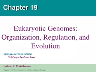
Chapter 19 EukaryoticGenomes: Organization, Regulation, and Evolution
Figure 19.1 Overview: How Eukaryotic Genomes Work and Evolve • In eukaryotes, the DNA-protein complex, called chromatin • Is ordered into higher structural levels than the DNA-protein complex in prokaryotes. How can this structure be ordered in this intricate manner?
Both prokaryotes and eukaryotes • Must alter their patterns of gene expression in response to changes in environmental conditions
Concept 19.1: Chromatin structure is based on successive levels of DNA packing • Eukaryotic DNA • Is precisely combined with a large amount of protein with the resulting chromatin undergoes striking changes during the cell cycle • When the cell prepare to mitosis, its chromatin coils and folds to form the chromosomes • Eukaryotic chromosomes • Contain an enormous amount of DNA contain a single linear DNA double helix that averages 200 million base pairs in humans
Nucleosomes, or “Beads on a String” • Proteins called histones • Are responsible for the first level of DNA packing in chromatin • Bind tightly to DNA because they have a high proportion of positively charged a.a that binds to the negatively charged DNA. • The association of DNA and histones • Seems to remain intact throughout the cell cycle
2 nm DNA double helix Histone tails His- tones 10 nm Histone H1 Linker DNA (“string”) Nucleosome (“bad”) (a) Nucleosomes (10-nm fiber) In electron micrographs • Unfolded chromatin has the appearance of beads on a string • Each “bead” is a nucleosome; The basic unit of DNA packing • Histones leave DNA only transiently during DNA replication but stay with DNA during transcription. Figure 19.2 a
30 nm Nucleosome (b) 30-nm fiber Higher Levels of DNA Packing • With the aid of the histone H1 (one of 5 histones involved in the coiling process), the beaded string coils to from a fiber roughly 30 nm in thickness known as the 30-nm chromatin fiber. Figure 19.2 b
Protein scaffold Loops Scaffold 300 nm (c) Looped domains (300-nm fiber) • The 30-nm fiber, in turn • Forms looped domains, making up a 300-nm fiber which are attached to a chromosome scaffold (platform) made of non-histone proteins. Figure 19.2 c
700 nm 1,400 nm (d) Metaphase chromosome • In a mitotic chromosome • The looped domains themselves coil and fold further compacting the chromatin forming the characteristic metaphase chromosome Figure 19.2 d
In interphase • Some portions of certain chromosomes exist in a very condensed state that can be seen under light microscope. This is called heterochromatin and is distinguished from the less compact chromatin that is called euchromatin.
Concept 19.2: Gene expression can be regulated at any stage, but the key step is transcription • All organisms • Must regulate which genes are expressed at any given time. i.e not every gene will be active at all times. • During development of a multicellular organism • Its cells undergo a process of specialization in form and function called cell differentiation. This results in several cell types reaching up to 200 different cells types in adult humans.
Differential Gene Expression • Each cell of a multicellular eukaryote • Expresses only a fraction of its genes (probably 20% ) at any given time. • In each type of differentiated cell • A unique subset of genes is expressed with the highly differentiated cells such as muscle cells expressing a lower number of genes at any given time. • Differences between cell types are NOT due to different genes but to different gene expression by cells with the same genome.
The genome of eukaryotes may contain tens of thousands of genes but only a small amount of DNA (1.5% in humans ) codes for proteins. • What determine which genes to be expressed? • Environmental signals, certain genes turn on while others turn off. • Cell differentiation during organism development. • The enzymes that transcribe DNA must locate the target genes at the right time. This task is like finding a needle in haystack. • When gene expression goes wrong, certain disease such as cancer can arise.
Signal NUCLEUS Chromatin Chromatin modification: DNA unpacking involving histone acetylation and DNA demethlation DNA Gene available for transcription Gene Transcription Exon RNA Primary transcript Intron RNA processing Tail mRNA in nucleus Cap Transport to cytoplasm CYTOPLASM mRNA in cytoplasm Degradation of mRNA Translation Polypetide Cleavage Chemical modification Transport to cellular destination Active protein Degradation of protein Degraded protein Figure 19.3 Many key stages of gene expression Stages in Gene Expression that can be regulated in eukaryotic cells; The colored boxes indicates these steps.
Regulation of Chromatin Structure • Genes within highly packed heterochromatin • Are usually not expressed
Chromatin changes Transcription RNA processing Translation mRNA degradation Protein processing and degradation Histone tails DNA double helix Amino acids available for chemical modification Histone Modification • Chemical modification of histone tails • Can affect the configuration of chromatin and thus gene expression Figure 19.4a (a) Histone tails protrude outward from a nucleosome
Acetylated histones Unacetylated histones (b) Acetylation of histone tails promotes loose chromatin structure that permits transcription Figure 19.4 b Histone acetylation • is the attachment of acetyl groups (--COCH3) to certain amino acids of histone proteins. • Seems to loosen chromatin structure and thereby lose their grip on DNA thus enhance transcription
DNA Methylation • Addition of methyl groups (…CH3) to certain bases in DNA • Genes are usually more highly methylated when they are not expressed. • De-methylation to certain inactive genes turns them on. • In some species DNA methylation guarantees long term inactivation of certain genes • Once methylated, genes stay as such through successive cell division. This property accounts for genomic imprinting in mammals where methylation permanently turns off either maternal or paternal allele of a certain gene.
Epigenetic Inheritance • Is the inheritance of traits transmitted by mechanisms not directly involving the nucleotide sequence. • Chromatin modifying enzymes are integral part of this process.
Regulation of Transcription Initiation • Chromatin-modifying enzymes provide initial control of gene expression • By making a region of DNA either more or less able to bind the transcription machinery. • Once a gene is optimally modified for expression, the initiation of transcription process is the most important.
Proximal control elements Enhancer (distal control elements) Poly-A signal sequence Termination region Exon Intron Intron Exon Exon DNA Downstream Upstream Promoter Transcription Poly-A signal Cleared 3 end of primary transport Primary RNA transcript (pre-mRNA) Exon Exon Intron Intron Exon 5 Chromatin changes RNA processing: Cap and tail added; introns excised and exons spliced together Transcription Intron RNA RNA processing Coding segment mRNA degradation Translation mRNA P Protein processing and degradation G P P Start codon Poly-A tail Stop codon 3 UTR (untranslated region) 5 Cap 5 UTR (untranslated region) Organization of a Typical Eukaryotic Gene • Associated with most eukaryotic genes are multiple control elements which are; • Segments of noncoding DNA that help regulate transcription by binding certain proteins Figure 19.5
The Roles of Transcription Factors • To initiate transcription • Eukaryotic RNA polymerase requires the assistance of proteins called transcription factors. These factors are required for all types of protein coding genes thus called general transcription factors. • One of these transcription factors recognize a DNA sequence called the TATA box within the promoter, while the others recognize other proteins including the RNA polymerase.
Enhancers and Specific Transcription Factors • The interaction of these transcription factors and RNA polymerase II with a promoter usually initiates transcription but inefficiently producing few RNA transcripts. • Control elements, proximal control elements (near the promoter) greatly improve the efficiency of promoters by binding additional transcription factors. • The more distant “ distal control elements” are called the enhancers which might be thousands of nucleotides away from the promoter or may be located in an intron.
Distal control element Promoter Activators Gene TATA box Enhancer General transcription factors Activator proteins bind to distal control elements grouped as an enhancer in the DNA. This enhancer has three binding sites. 1 DNA-bending protein Group of Mediator proteins A DNA-bending protein brings the bound activators closer to the promoter. Other transcription factors, mediator proteins, and RNA polymerase are nearby. 2 RNA Polymerase II Chromatin changes The activators bind to certain general transcription factors and mediator proteins, helping them form an active transcription initiation complex on the promoter. 3 Transcription RNA Polymerase II RNA processing mRNA degradation Translation Protein processing and degradation Transcription Initiation complex RNA synthesis • An activator • Is a protein that binds to an enhancer and stimulates transcription of a gene Figure 19.6
Some specific transcription factors function as repressors • To inhibit expression of a particular gene • Some activators and repressors • Act indirectly by influencing chromatin structure • A gene present in the region of chromatin with high levels of histone acetylation is able to bind to the transcription machinery while the one with low levels can not bind.
In yeast and some mammals some activator recruit proteins that acetylate histones near the promorter of a gene thus enhance transcription. Repressors recruit proteins that deacetylate histones leading to reduced transcription. This phenomenon is called gene silencing.
Coordinately Controlled Genes • Unlike the genes of a prokaryotic operon which are regulated by one promoter • Coordinately controlledeukaryotic genes each have a promoter and control elements even if they are located close to each other on the same chromosome. • The same regulatory sequences • Are common to all the genes of a group, enabling recognition by the same specific transcription factors • An example of such coordinate control is the activation of a variety of genes by steroid home (sex hormones). • Genes with the same control elements are activated by the same chemical signals.
Mechanisms of Post-Transcriptional Regulation • The expression of a protein-coding gene is ultimately measured in terms of amount of functional protein the cell makes. • In theory a gene expression may be blocked or stimulated at any post-transcriptional step. • A cell can therefore, use its regulatory mechanisms that operate after the transcription to fine tune gene expression in response to environmental changes without altering its transcription patterns.
Chromatin changes Transcription RNA processing mRNA degradation Translation Protein processing and degradation Exons DNA Primary RNA transcript RNA splicing or mRNA RNA Processing • In alternative RNA splicing • Different mRNA molecules are produced from the same primary transcript, depending on which RNA segments are treated as exons and which as introns. Figure 19.8
mRNA Degradation • The life span of mRNA molecules in the cytoplasm • Is an important factor in determining the protein synthesis in a cell • Prokaryotic mRNA have a very short life span i.e they are degraded enzymatically after few minutes. • In contrast eukaryotic mRNA life span is typically hours, but could be days or even weeks such mRNA for hemoglobin polypeptides • It is believed that the removal of the 5’ cap provides a site for the nuclease enzymes to chew up the mRNA. • Nucleotide sequences that affect mRNA stability are normally found in the trailer region (un-translated region, UTR) at the 3’ end of the molecule.
Recent discoveries • RNA interference by single-stranded microRNAs (miRNAs) • Can lead to degradation of an mRNA or block its translation • This happen due to a small interfering RNAs (siRNAs) which the same size as miRNAs and does the same function 5 4 3 1 2 The miRNA-protein complex prevents gene expression either by degrading the target mRNA or by blocking its translation. One strand of each short double- stranded RNA is degraded; the other strand (miRNA) then associates with a complex of proteins. The bound miRNA can base pair with any target mRNA that contains the complementary sequence. The micro- RNA (miRNA) precursor folds back on itself, held together by hydrogen bonds. An enzyme called Dicer moves along the double- stranded RNA, cutting it into shorter segments. 5 Chromatin changes Transcription RNA processing Protein complex mRNA degradation Translation Protein processing and degradation Dicer Degradation of mRNA OR miRNA Target mRNA Blockage of translation Hydrogen bond Figure 19.9
Initiation of Translation • The initiation of translation of selected mRNAs • Can be blocked by regulatory proteins that bind to specific sequences or structures of the mRNA. This adds another opportunity for regulating gene expression • Alternatively, translation of all the mRNAs in a cell • May be regulated simultaneously. This global control usually involves activation or inactivation of one or more of the protein factors required to initiate translation.
Protein Processing and Degradation • The final opportunities for controlling gene expression occurs after translation • Various types of protein processing, including cleavage and the addition of chemical groups, or the length of time each protein functions in the cell are subject to control. Examples; • Cleaving pro-insulin to form the active form, insulin. • Addition of phosphate groups or sugars • Cyclins which are proteins involved in regulating the cell cylce must be short-lived if the cell to function properly.
Chromatin changes Transcription RNA processing Proteasome and ubiquitin to be recycled Ubiquitin Translation mRNA degradation Proteasome Protein processing and degradation Protein fragments (peptides) Protein to be degraded Ubiquinated protein Protein entering a proteasome Proteasomes • The protein molecules to be degraded must be bind to another protein called ubiquitin before being degraded by a giant protein complexes called proteasome. 3 1 2 Enzymatic components of the proteasome cut the protein into small peptides, which can be further degraded by other enzymes in the cytosol. Multiple ubiquitin mol- ecules are attached to a protein by enzymes in the cytosol. The ubiquitin-tagged protein is recognized by a proteasome, which unfolds the protein and sequesters it within a central cavity Figure 19.10
Concept 19.3: Cancer results from genetic changes that affect cell cycle control • The gene regulation systems that go wrong during cancer turn out to be the very same systems that play important roles in embryonic development
Types of Genes Associated with Cancer • The genes that normally regulate cell growth and division during the cell cycle • Include genes for growth factors, their receptors, and the intracellular molecules of signaling pathways. • Its is believed that many causing-cancer mutations results from environmental influences such as chemical carcinogens, UV light, X-Ray or certain viruses.
Examples of viruses that cause cancer are; • EBV that cause infectious mononucleosis and is associated with Burkitt’s lymphoma • Papiloma virus associated with cervix cancer • HTLV-1 virus associated with some adults leukemia All tumor viruses transform cells into cancer cells by integrating their nucleic acids with host DNA.
Oncogenes and Proto-Oncogenes • Oncogenes • Are cancer-causing genes • Proto-oncogenes • Are normal cellular genes that code for proteins that stimulate normal cell growth and division
How a proto-oncogene become an oncogene? • An oncogene arises from a genetic change that leads to an increase in either; • the amount of the proto-oncogene’s protein product • or the intrinsic activity of each protein molecule. • Now these genetic changes fall into three catergories; (Figure 19-11).
Gentic changes that change proto-onco to oncogene • movement of DNA within the genome, where it might land close to an especially active promoter that increase transcription of the gene making it an oncogene. • amplification of proto-oncogene which increase the number of copies of oncogene inside the cell • point mutation in a proto-oncogene that change the gene’s protein product to a more active or more resistant to degradation than the normal protein.
Proto-oncogene DNA Translocation or transposition: gene moved to new locus, under new controls Point mutation within a control element Point mutation within the gene Gene amplification: multiple copies of the gene New promoter Oncogene Oncogene Hyperactive or degradation- resistant protein Normal growth-stimulating protein in excess Normal growth-stimulating protein in excess Normal growth-stimulating protein in excess • A DNA change that makes a proto-oncogene excessively active • Converts it to an oncogene, which may promote excessive cell division and cancer Figure 19.11
Tumor-Suppressor Genes • Tumor-suppressor genes • Encode proteins that inhibit abnormal cell division. • Protein products of tumor suppressor genes could be; • proteins normally repair damaged DNA • some proteins control adhesion of cells to each other or to an extracellular matrix • other proteins are component of signaling pathways that inhibit the cell cycle.
Interference with Normal Cell-Signaling Pathways • Many proto-oncogenes and tumor suppressor genes • Encode components of growth-stimulating and growth-inhibiting pathways, respectively. • What protein products of cancer genes do or fail to do? • Let us look at products of two cancer genes; the ras proto-ocogenes and p53 tumor suppressor gene.
Mutations in these genes are very common in human cancers; ras is mutated in about 30% of human cancers while it is 50% for p53. • Both proteins, ras and p53 are parts of the signal transduction pathways that convey external signal to the DNA in the nucleus. Figure 19-12 shows two possibilities of an outcome when either gene goes wrong; • one an over expression of ras protein that ends in the increase in the production of protein that stimulates cell cycle and leads to cancer • An under expression of a protein that inhibits the cell cycle as a result of mutation in the p53 gene and leads to cancer.
The p53 gene is names as it encodes for a protein of 53 kDa molecular weight. It is very important to the point that its name is “the guardian angel of the genome”. How does the p53 works in protecting the cells form cancer? • A damage to the cell’s DNA leads to the expression of p53 • This in turn lead to the activation of a p21gene whose product leads to halting the cell cycle thus allowing time for the cell to repair its damaged DNA • The protein p53 can turn on genes directly involved in DNA repair • When DNA damage is irreparable, p53 protein activates a suicide genes whose protein products cause cell death in a process called apoptosis
Cell cycle stimulating pathway • This pathway is triggered by a growth factor that binds to its receptor in the plasma membrane. • The signal is relayed to a G protein called Ras. Like all G proteins, Ras is active when GTP is bound to it. • Ras passes the signal to a series of protein kinases. • The last kinase activates a transcription activator that turns on one or more genes. • For proteins that stimulate the cell cycle. If a mutation makes Ras or any other pathway component abnormally active, excessive cell division and cancer may result. See next slide for explanation.
1Growth factor G protein Protein kinases (phosphorylation cascade) Receptor Transcription factor (activator) 2 5 3 4 • The Ras protein, encoded by the ras gene • Is a G protein that relays a signal from a growth factor receptor on the plasma membrane to a cascade of protein kinases Mutation Hyperactive Ras protein (product of oncogene) issues signals on its own Nucleus Gene expression Protein that Stimulates the cell cycle
Cell cycle inhibiting pathway • In this pathway, DNA damage is an intracellular signal that is passed via protein kinases and leads to activation of p53. • Activated p53 promotes transcription of the gene for a • protein that inhibits the cell cycle. • The resulting suppression of cell division ensures that the damaged DNA is not replicated. • Mutations causing deficiencies in any pathway component can contribute to the development of cancer. • See next slide for explanation.
Protein kinases 2 Active form of p53 3 DNA damage in genome 1 • The p53 gene encodes a tumor-suppressor protein • That is a specific transcription factor that promotes the synthesis of cell cycle–inhibiting proteins MUTATION UV light Defective or missing Transcription factor, such as p53, cannot activate transcription DNA Protein that inhibits the cell cycle Figure 19.12b